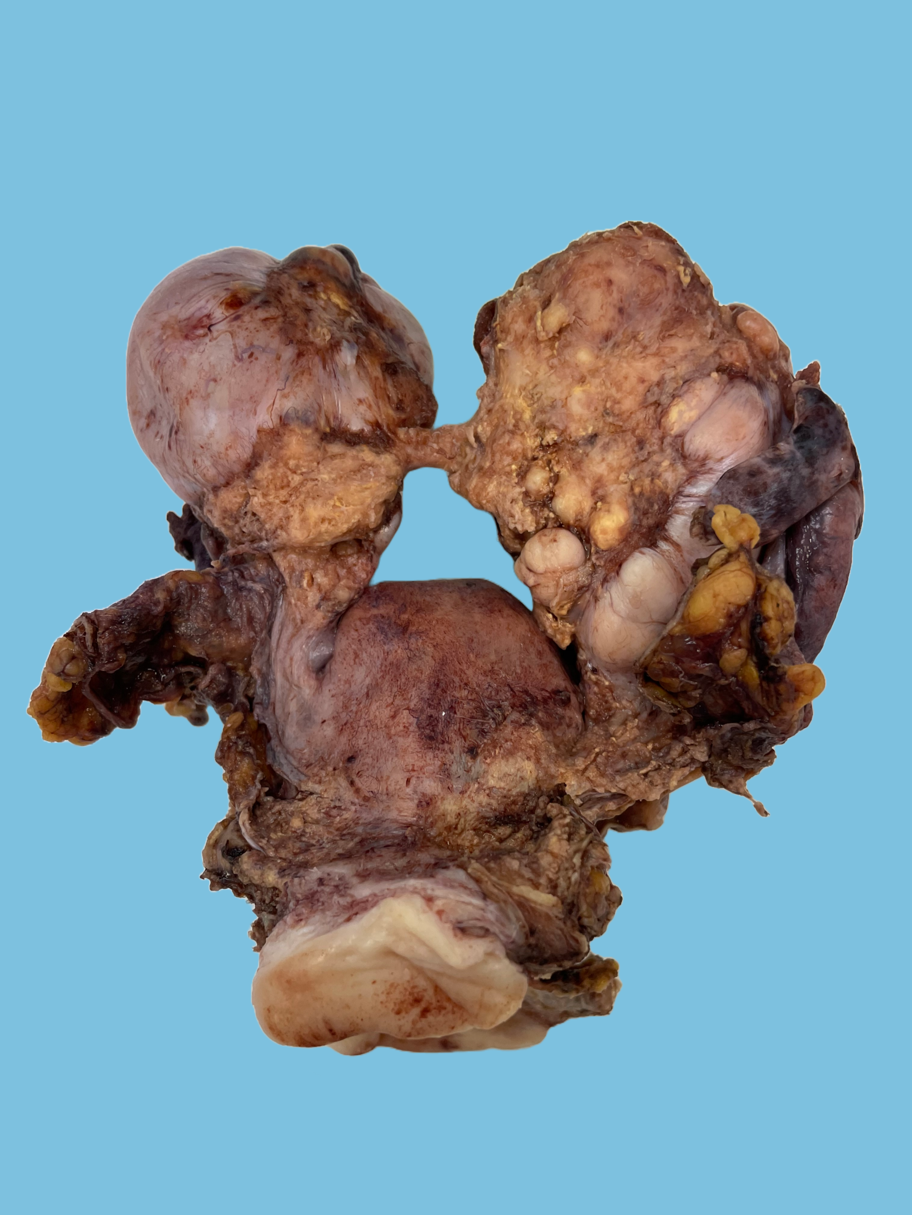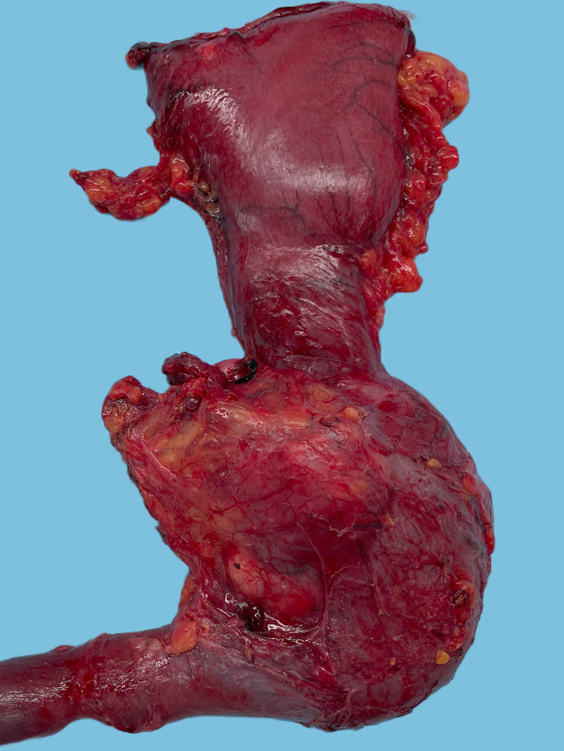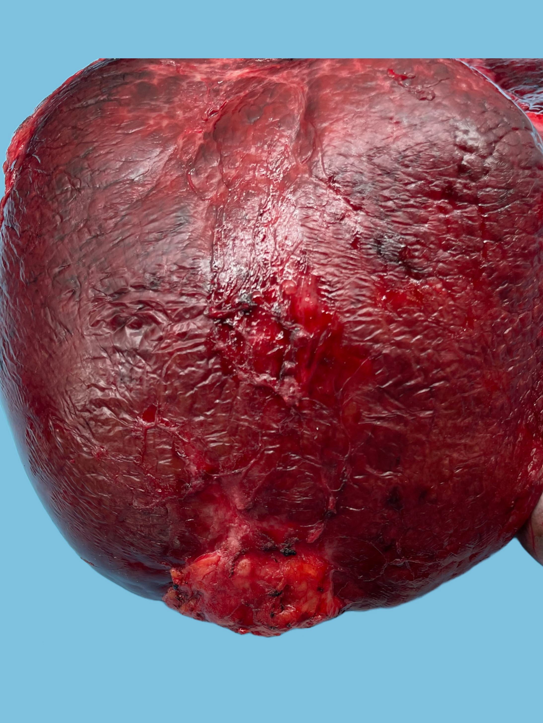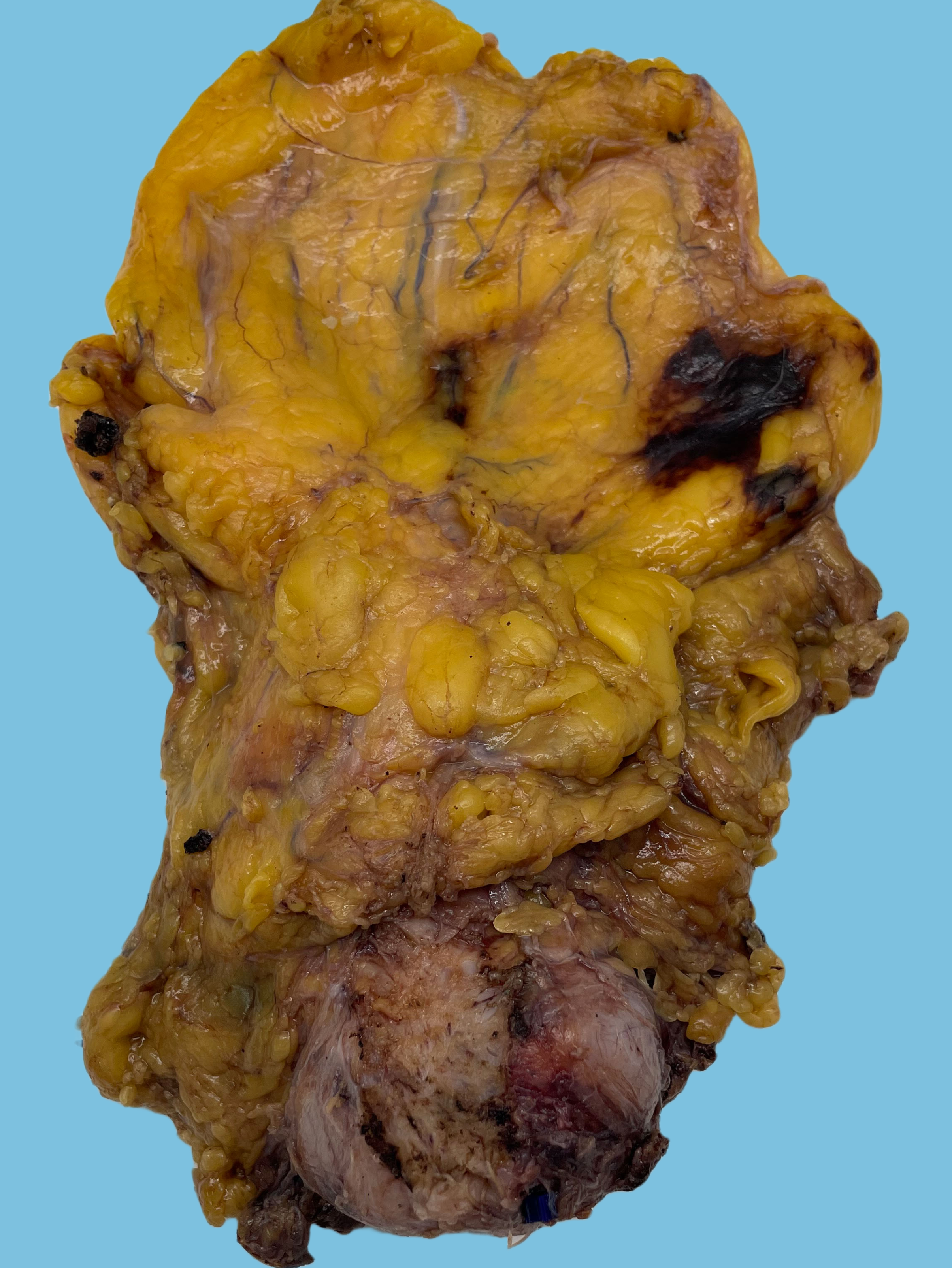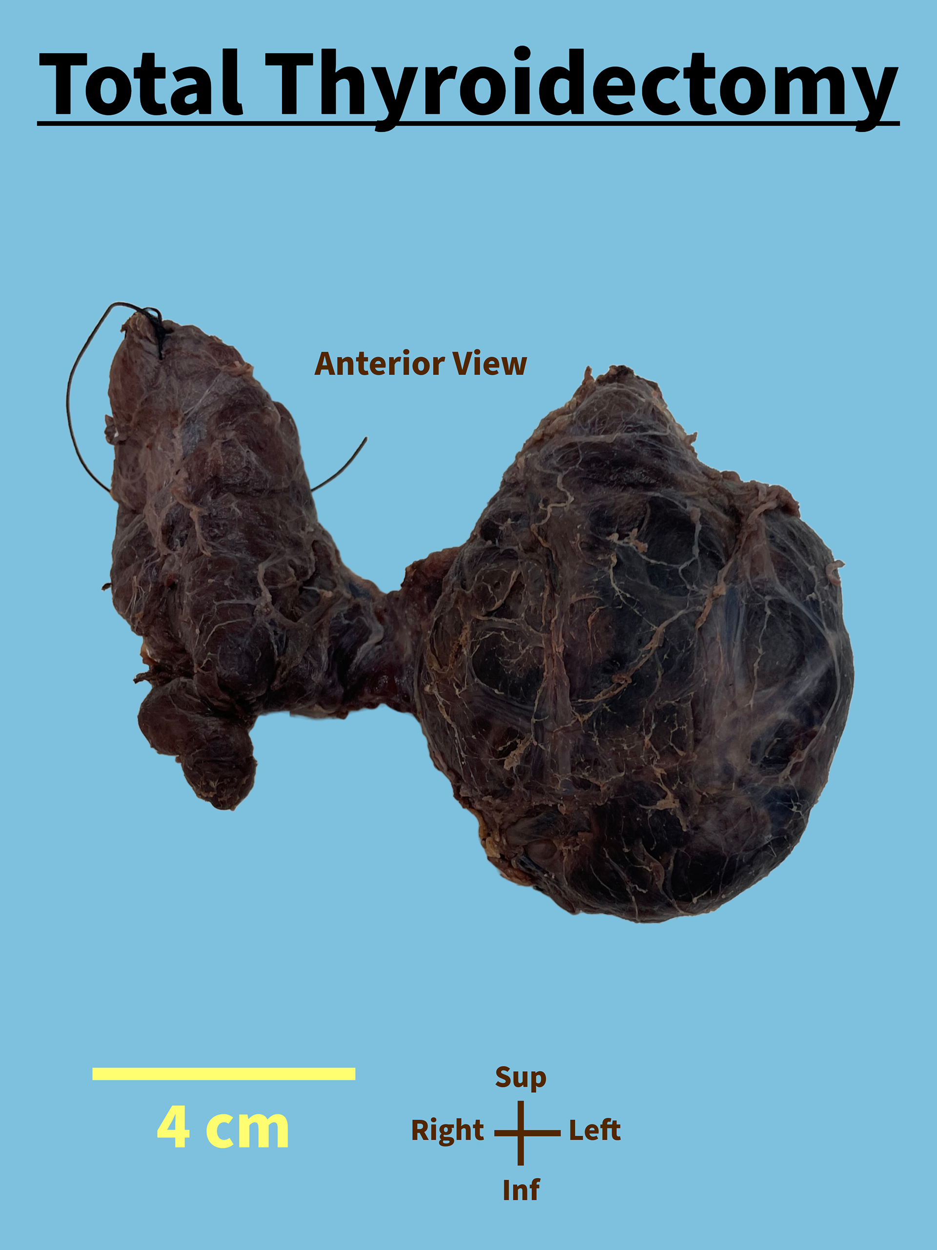
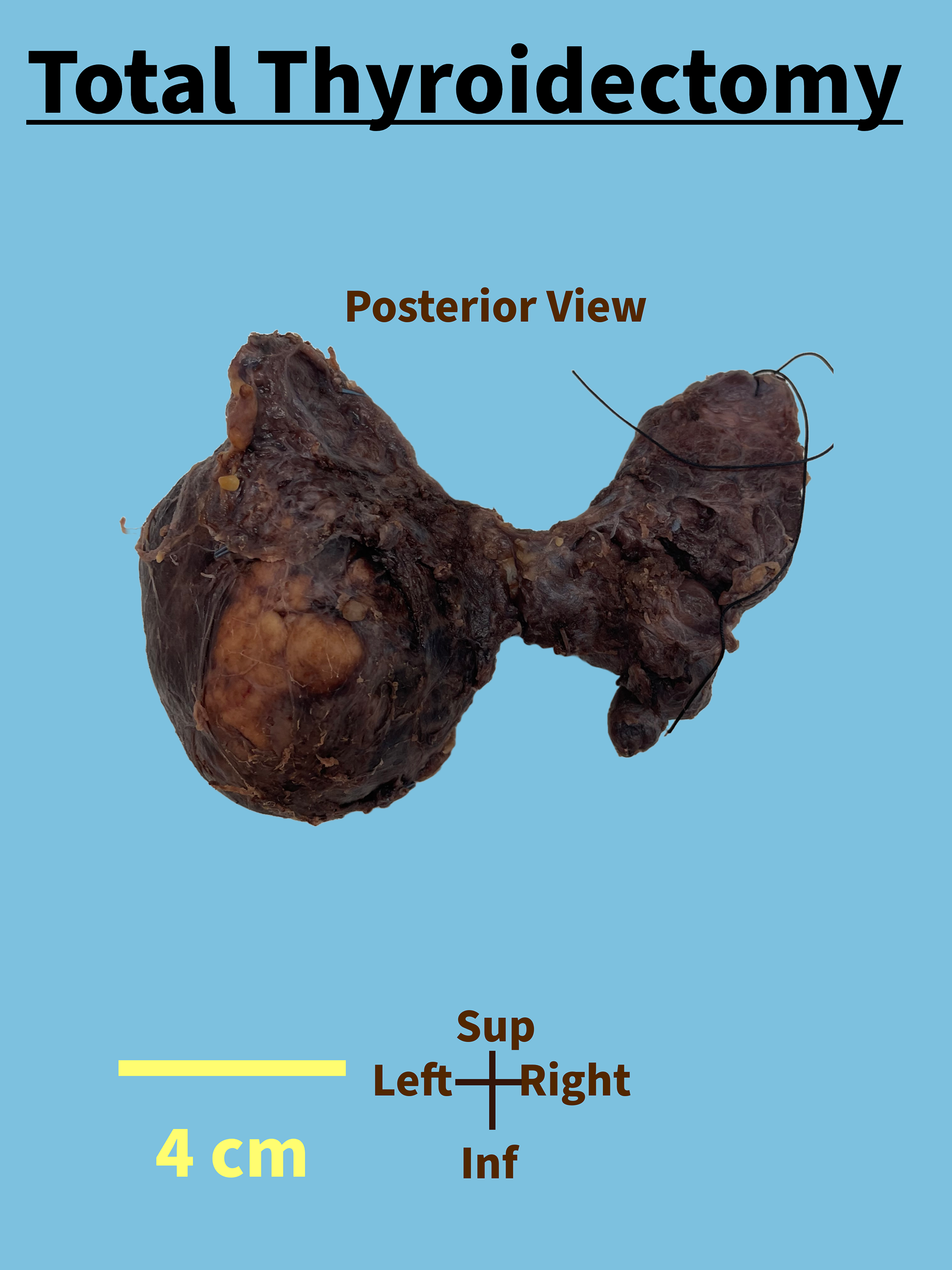
Received fresh for routine processing and subsequently placed in formalin by the Surgical Pathology Department, labeled with the patient's ID and labeled on the container " total thyroidectomy, suture marks right superior pole" is an oriented 116 g total thyroidectomy specimen with the left lobe (8.0 cm inferior-superior, 6.0 cm medial-lateral, 3.8 cm posterior-anterior), right lobe (7.0 cm inferior-superior, 3.0 cm medial-lateral, 1.9 cm posterior-anterior), and isthmus (1.5 x 1.5 x 0.3 cm). The right posterior surface is tan-brown, roughed and remarkable for a 3.0 x 1.1 cm tan-brown defect in the right lobe. The anterior surface is tan-brown and nodular.
The specimen is inked as follows: Posterior–black, right anterior–blue, left anterior–green, anterior isthmus–orange.
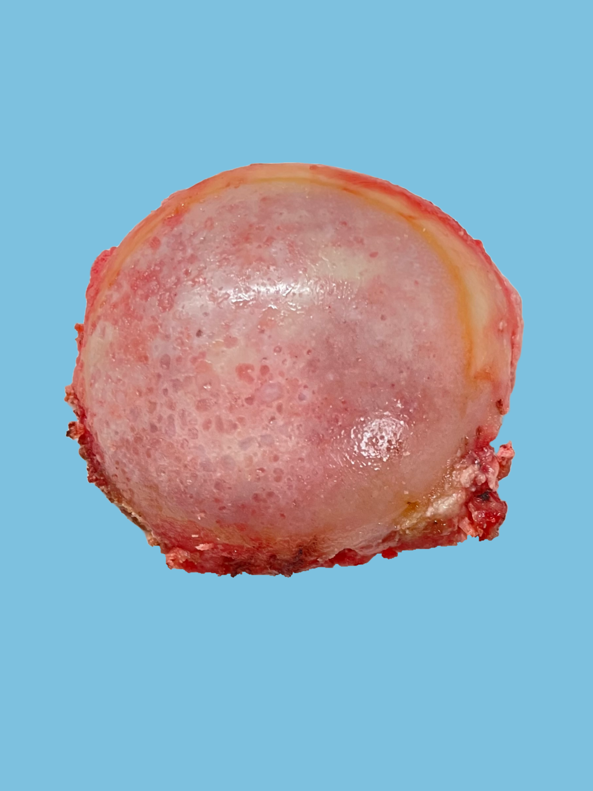
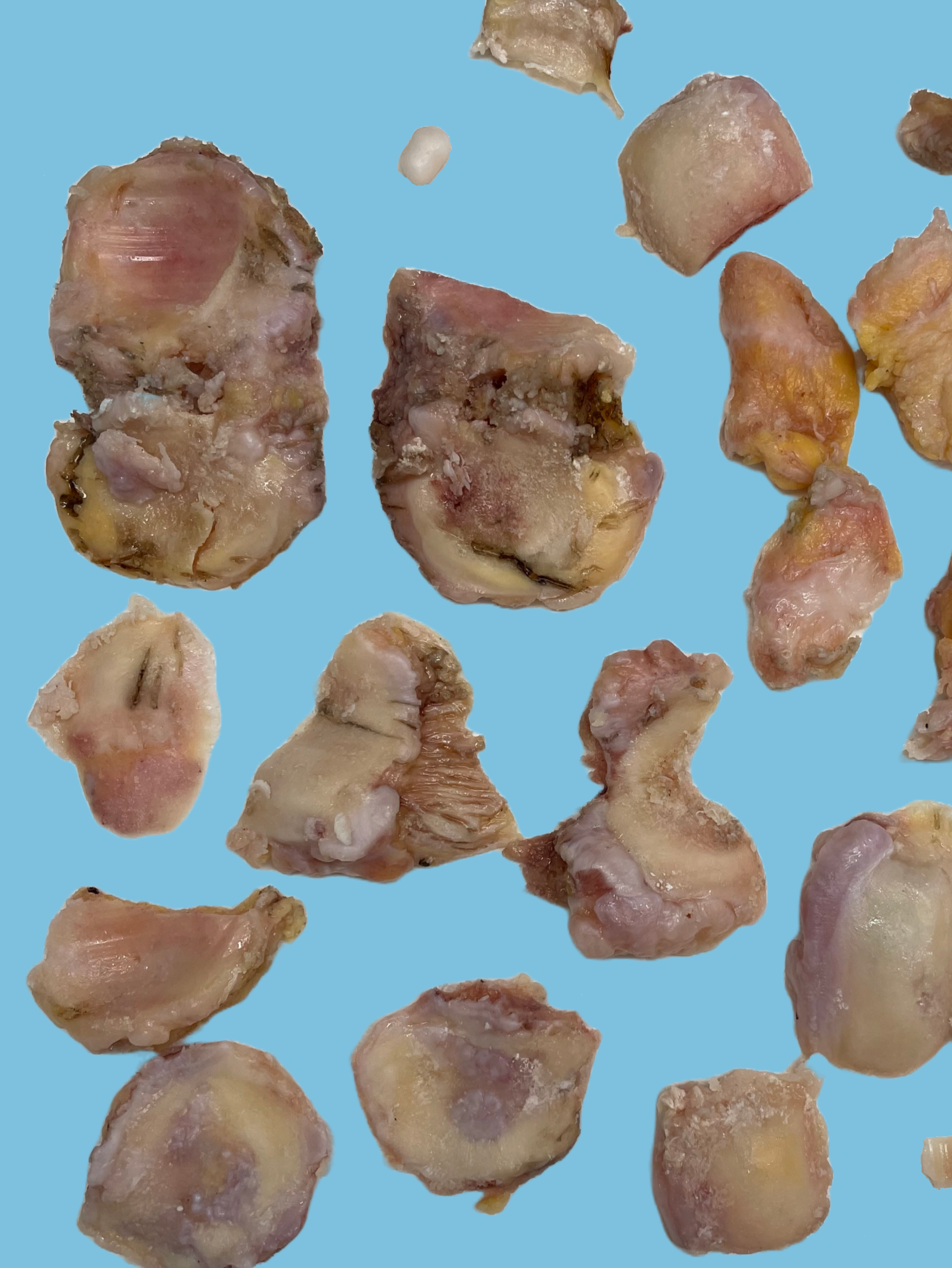
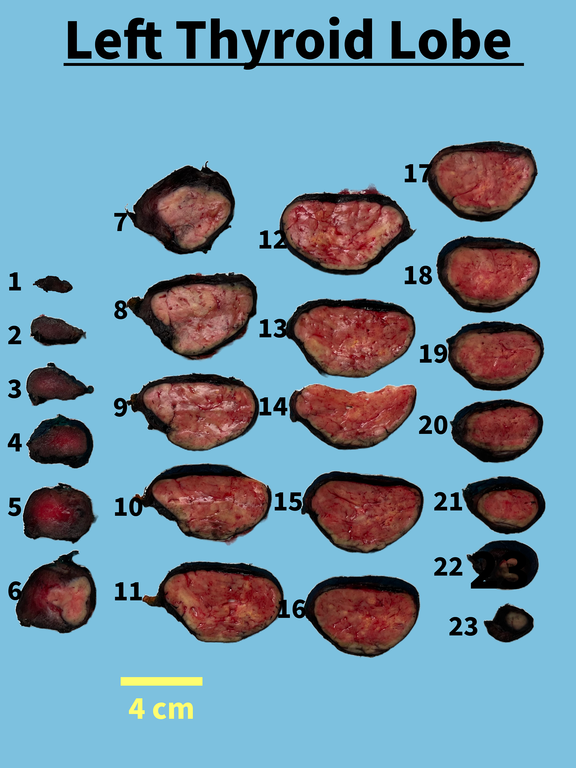
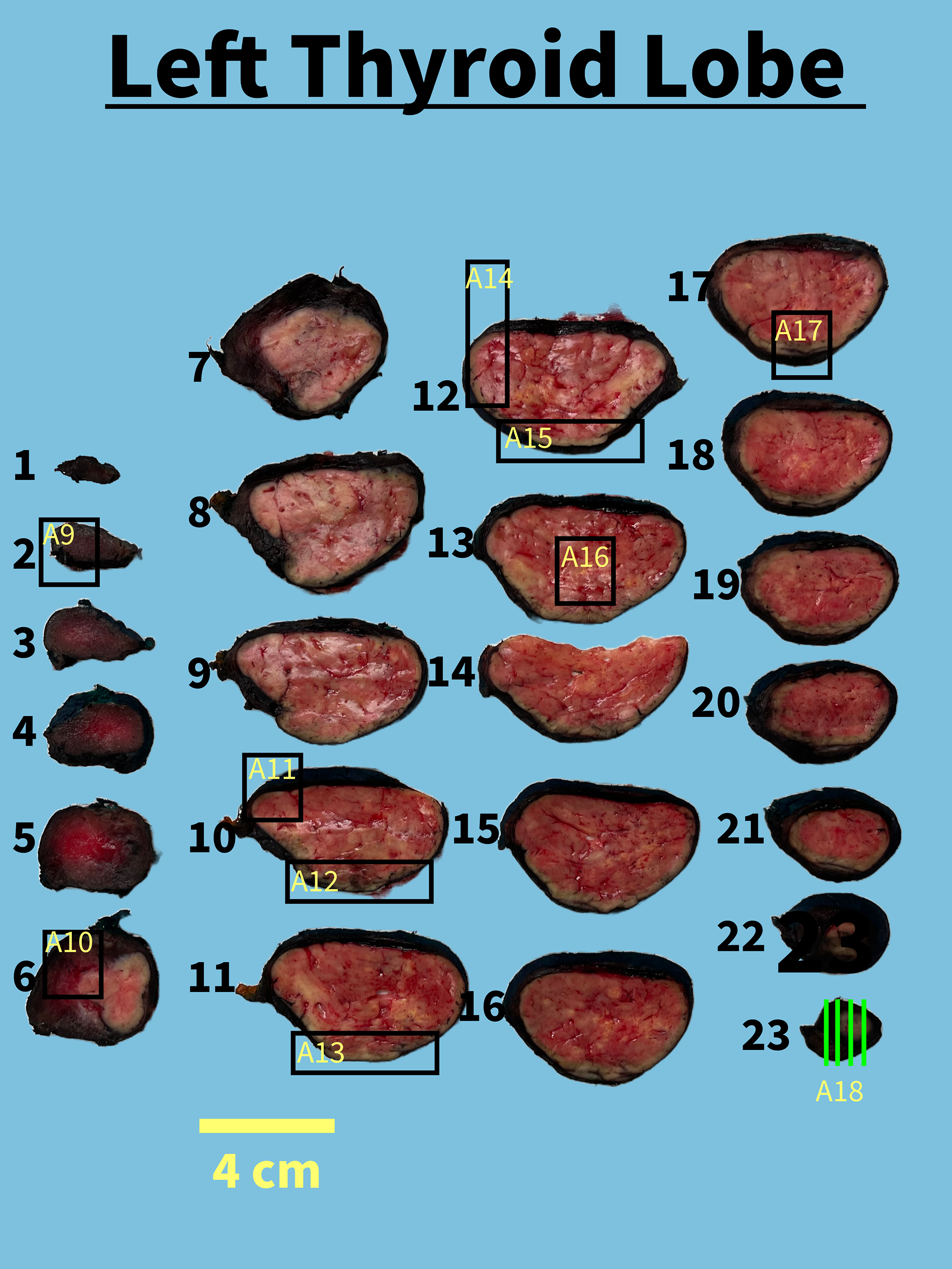
The left lobe is serially sectioned from superior to inferior into 23 slices to reveal a 6.4 x 6.2 x 3.6 cm tan-white well-circumscribed soft mass occupying the lower pole and mid pole. The mass abuts the posterior, anterior surfaces, is 0.4 cm from the isthmus and 1.9 cm from the right lobe. The remaining left lobe parenchyma is diffuse red-brown.
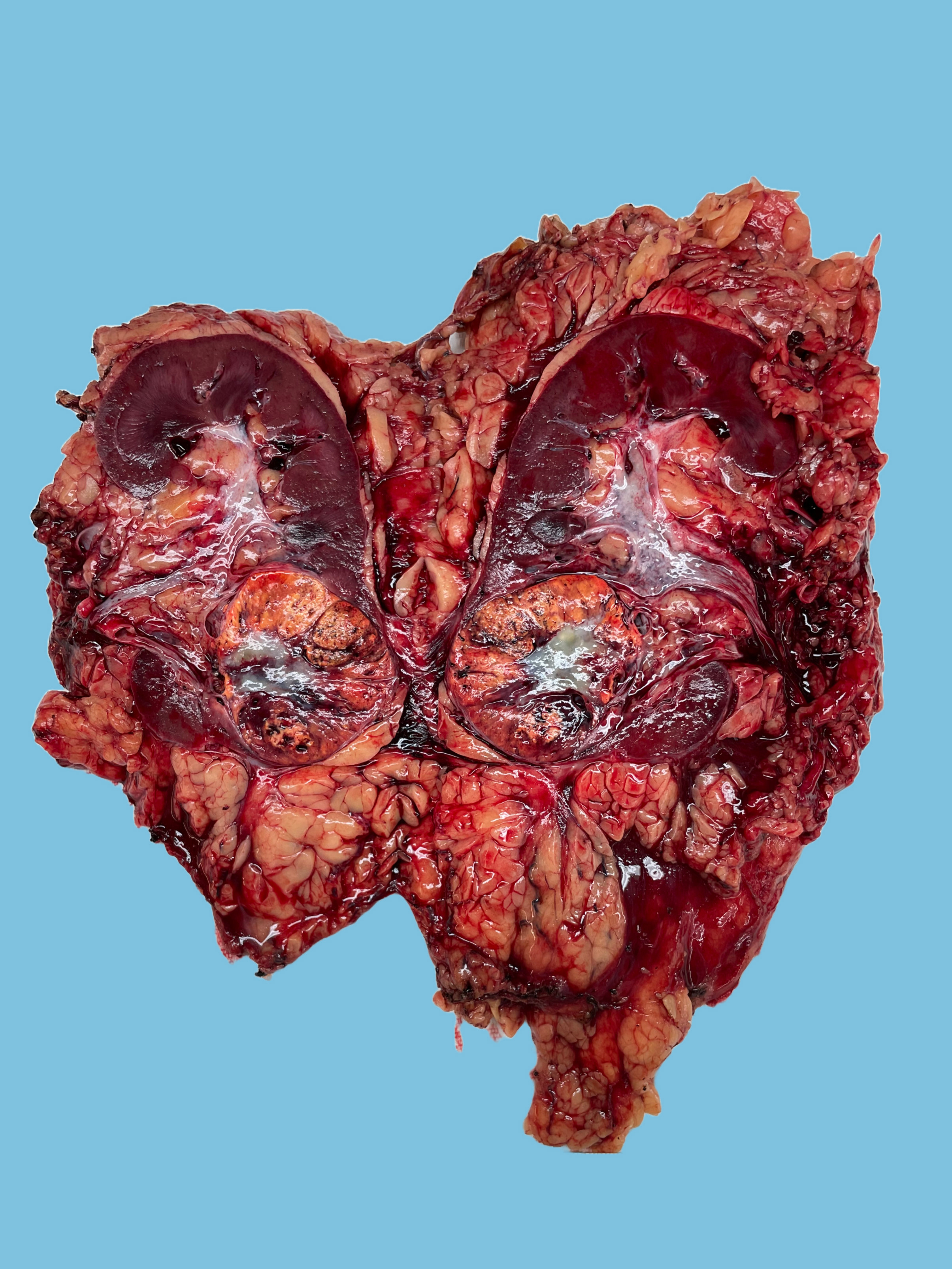
The right lobe and isthmus is serially sectioned from superior to inferior into 21 slices to reveal 1.8 x 1.4 x 1.2 cm encapsulated lesion within the lower pole in slices 13-17. The lesion abuts the defected area, is within 0.1 cm of the anterior surface, 0.5 cm from the posterior surface, is 1.7 cm from the isthmus and greater than 3.0 cm from the left lobe. Additionally identified are multiple white nodules ranging from 0.2 to 0.5 cm in greatest dimension diffusely throughout. The remaining parenchyma is diffuse red-brown.
Representative sections to include the entire encapsulated lesion are submitted as follows:
A1: Right lobe slice 4 entirely, superior pole parenchyma with nodule
A2: Right lobe slice 9, midpole parenchyma with nodule
A3: Right lobe slice 13, encapsulated lesion superior aspect, abutting defect
A4: Right lobe slice 14, encapsulated lesion
A5: Right lobe slice 15, encapsulated lesion to anterior surface
A6: Right lobe slice 16, encapsulated lesion to closest posterior surface
A7: Right lobe, slice 17, encapsulated lesion inferior aspect
A8: Right lobe, slice 19, lower pole parenchyma with nodule
A9: Left lobe slice 3, superior pole parenchyma
A10: Left lobe slice 6, mass to uninvolved
A11-A12: Left lobe slice 10
A11: Mass to close at the isthmus
A12: Mass abutting posterior-anterior, bisected
A13: Left lobe slice 11, mass to anterior surface, bisected
A14-A15: Left lobe slice 12
A14: Mass to anterior surface, bisected
A15: Mass to posterior surface, bisected
A16: Left lobe slice 13, central mass areas
A17: Left lobe slice 17, mass to posterior surface, bisected
A18: Left lobe slice 23, Inferior aspect of mass, perpendicular