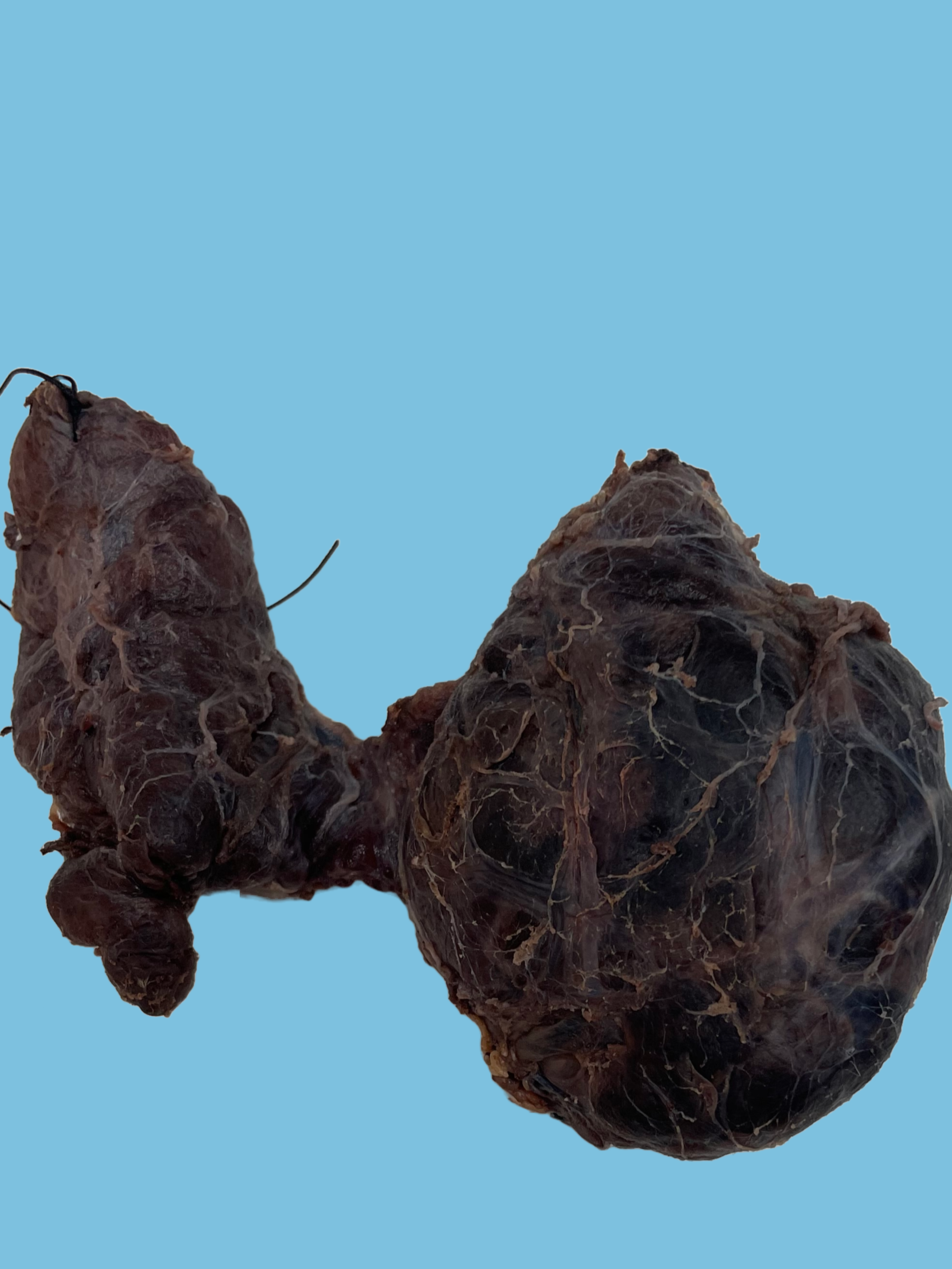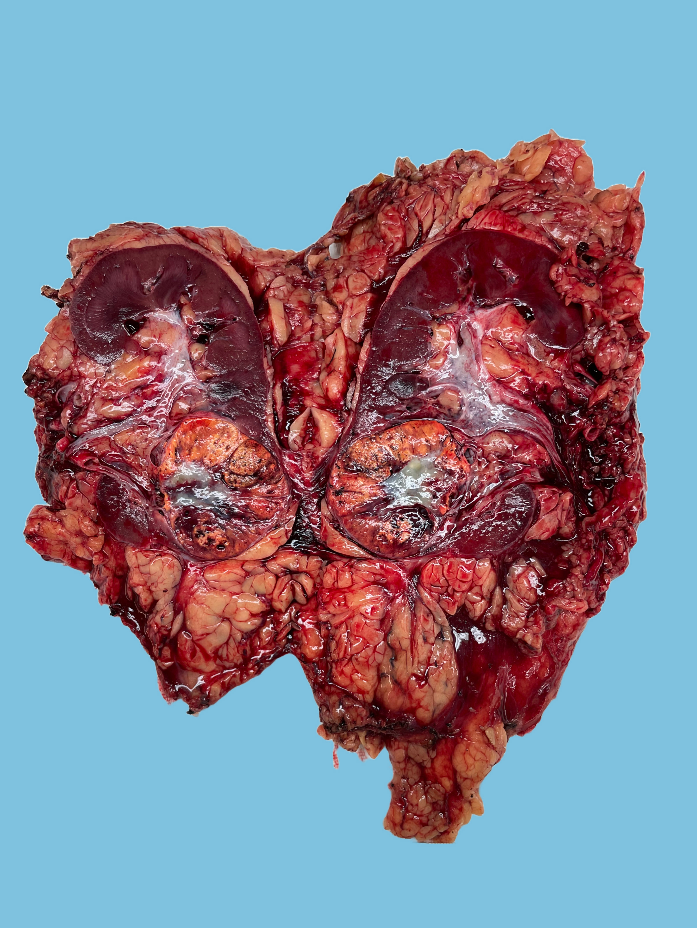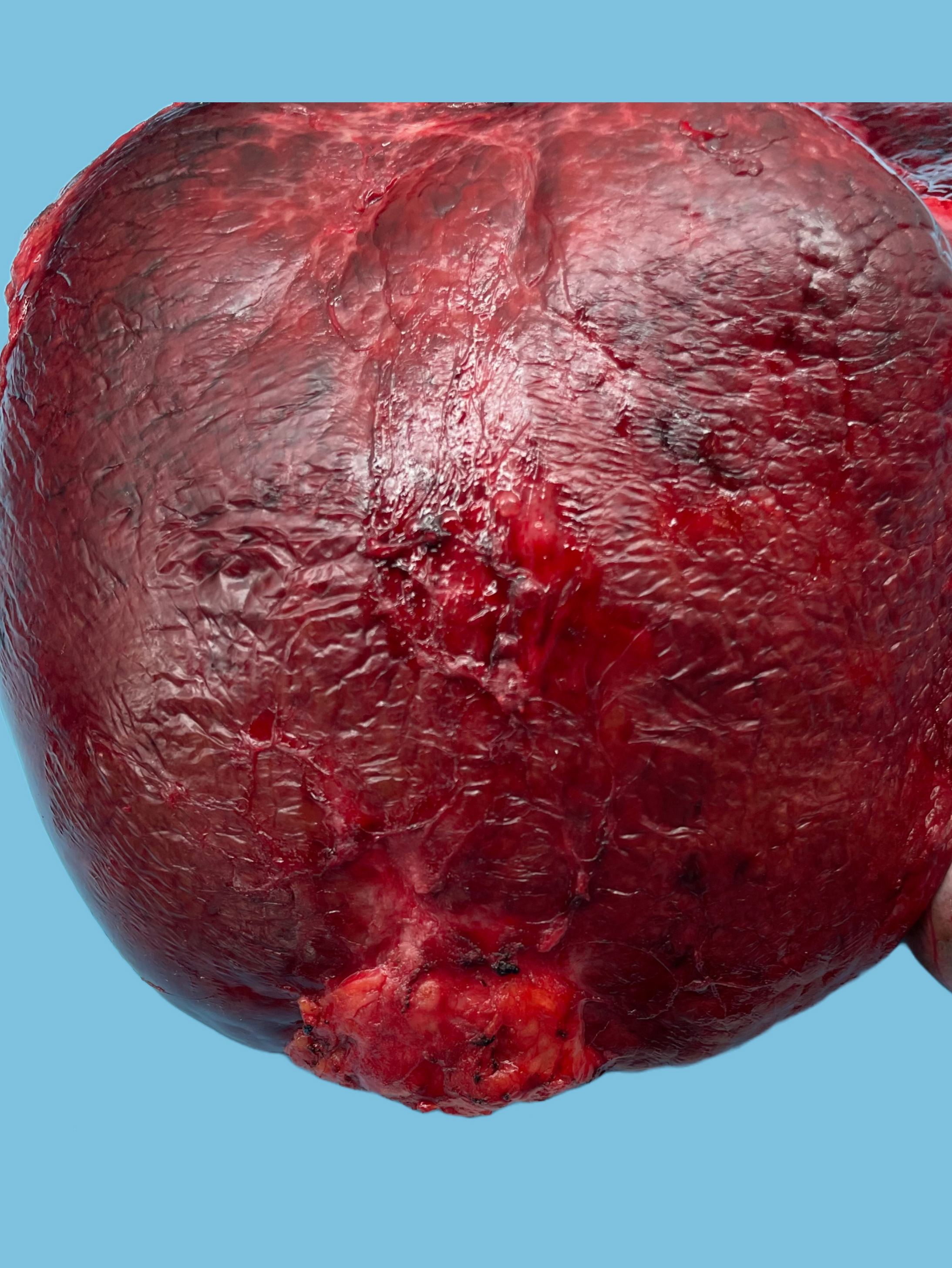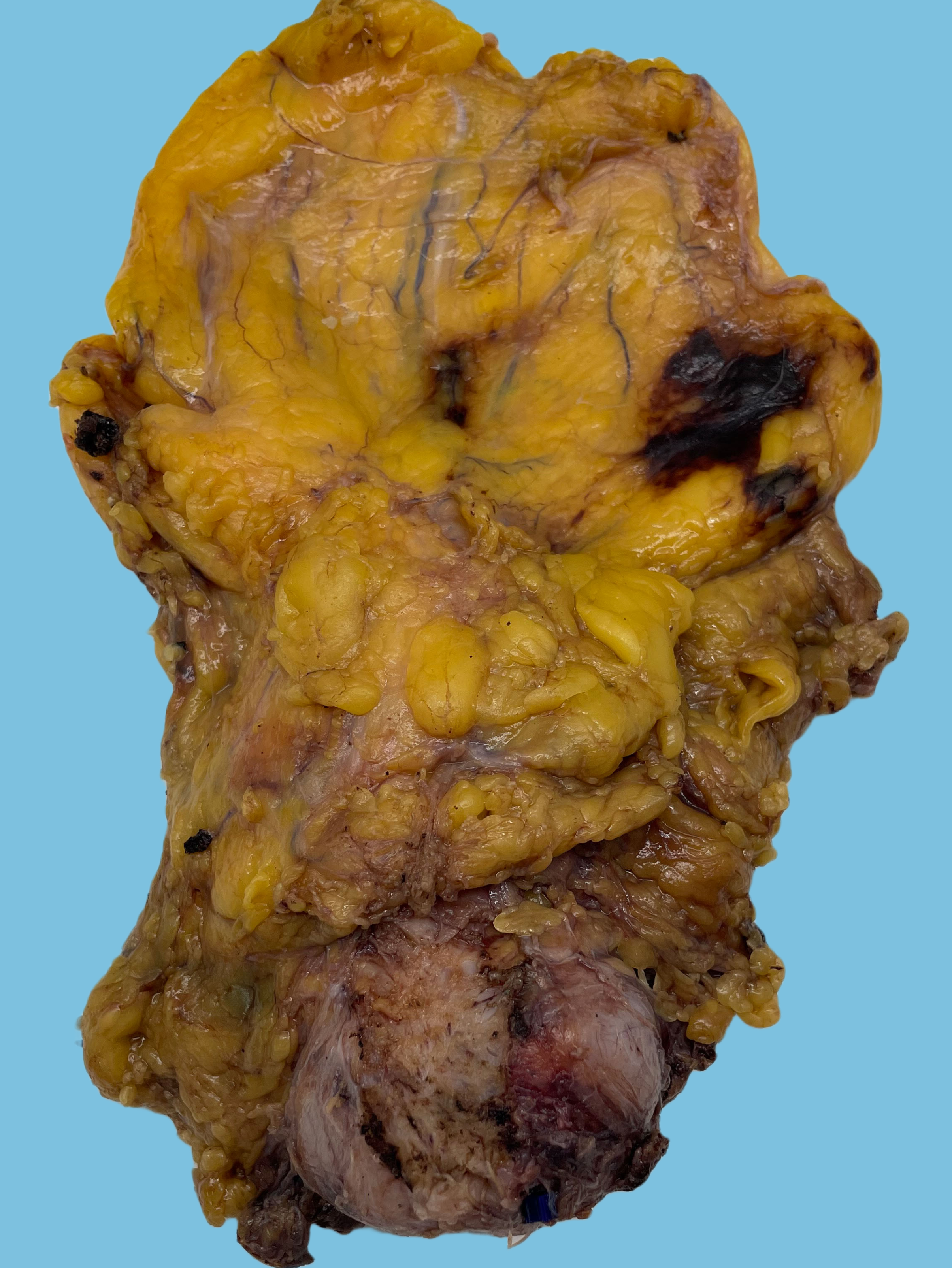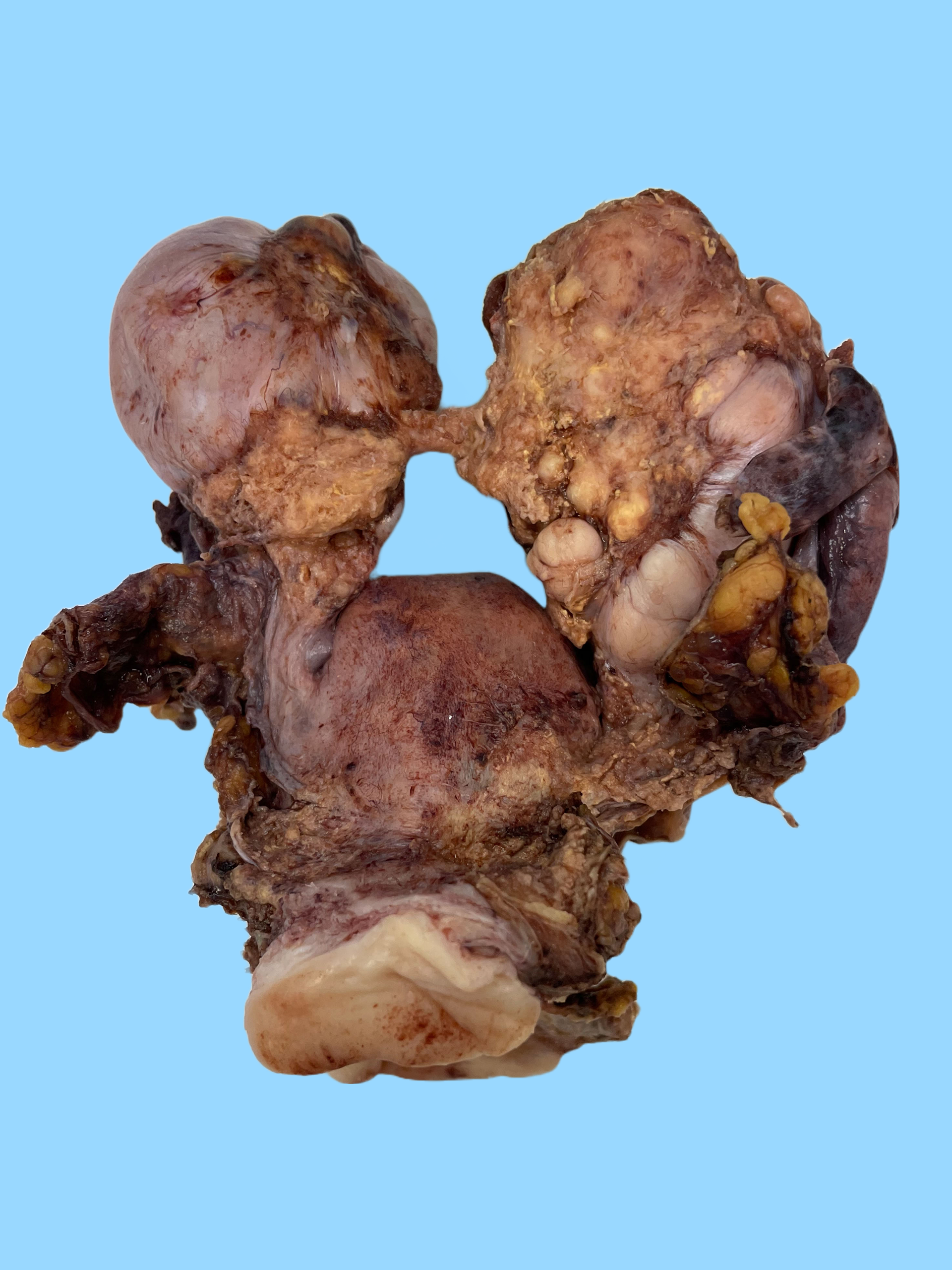
Received fresh labeled "uterus, cervix with bilateral ovaries and fallopian tube" is a 319 g hysterectomy specimen consisting of uterus, cervix, and attached bilateral adnexa. The right ovary (8.5 x 5.2 x 5.0 cm) has tan-white nodular external surface remarkable for a 6.0 x 5.5 cm tan-yellow white friable solid and cystic mass covering the entirety of the ovary external surface. The mass extends across to cover the entirety of left ovary. Additionally identified are areas of tan-yellow friable mass like areas in the right broad ligament, lower uterine serosa, and at both parametrium margins.
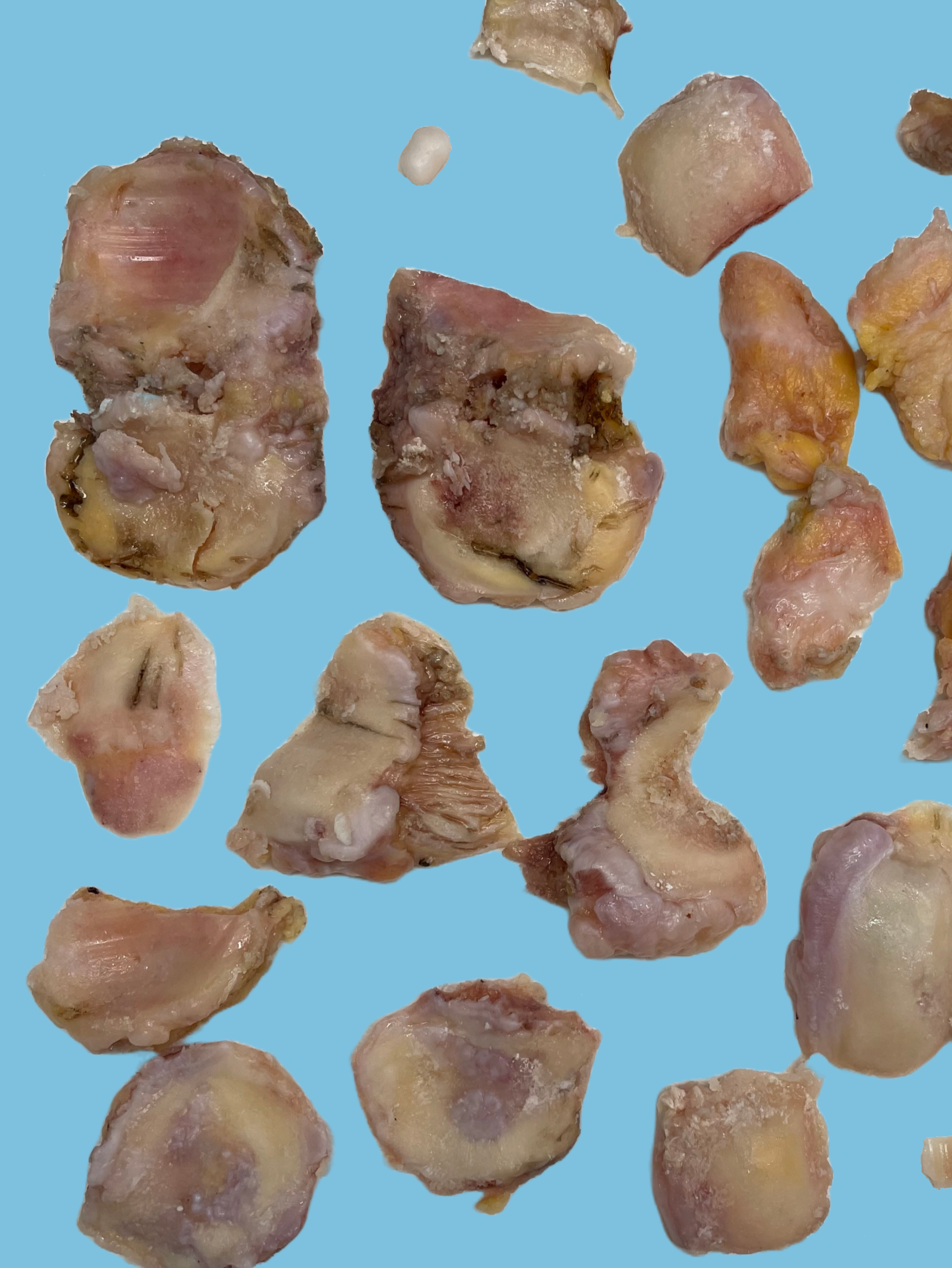
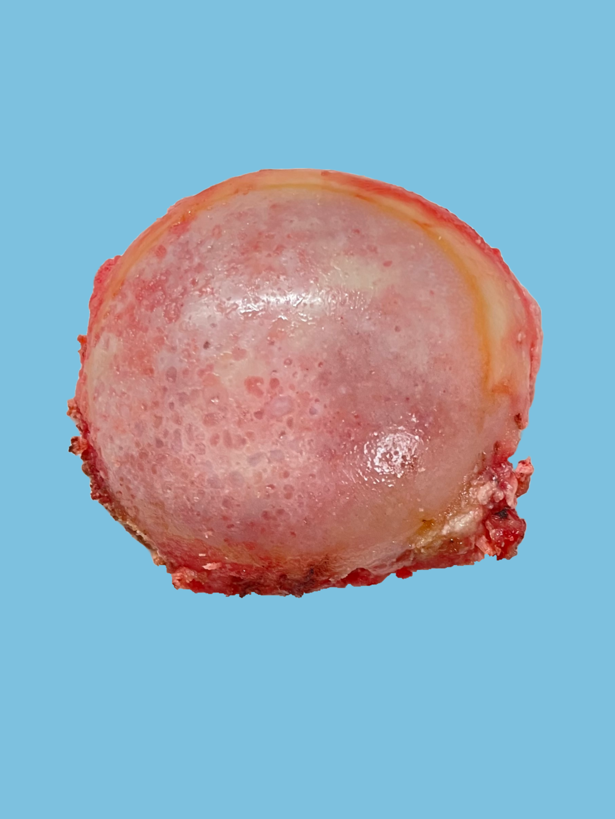
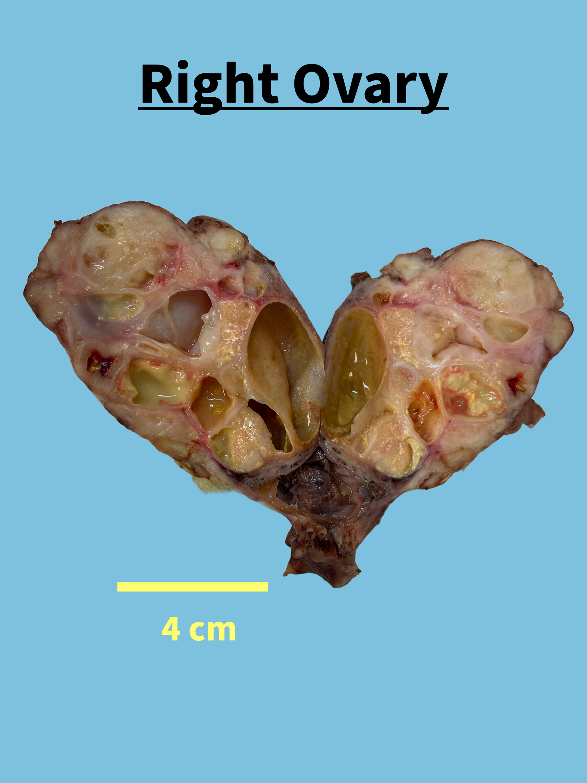
Sectioning of the right ovary reveals a 8.5 x 5.2 x 5.0 cm multiloculated solid-cystic mass. The mass is comprised of white firm nodules and cysts (ranging from 0.4 to 4.2 cm in greatest dimension) filled with yellow opaque fluid, and hemorrhagic/exudative contents. The cyst wall thickness measures up to 0.2 cm. The largest cyst's internal surface is remarkable for a 2.5 x 2.0 cm area of white papillary excrescences comprising 10 % of the overall specimen. No ovarian parenchyma is identified.
The right fimbriated fallopian tube (9.5 cm in length, diameter ranging from 0.4 to 1.0 cm) is intimately adherent to the right ovary, and has purple-tan serosa remarkable for multiple paratubal cysts ranging from 0.2 x 0.3 cm in greatest dimension with cloudy contents. Sectioning reveals a lumen ranging from pinpoint to stellate.
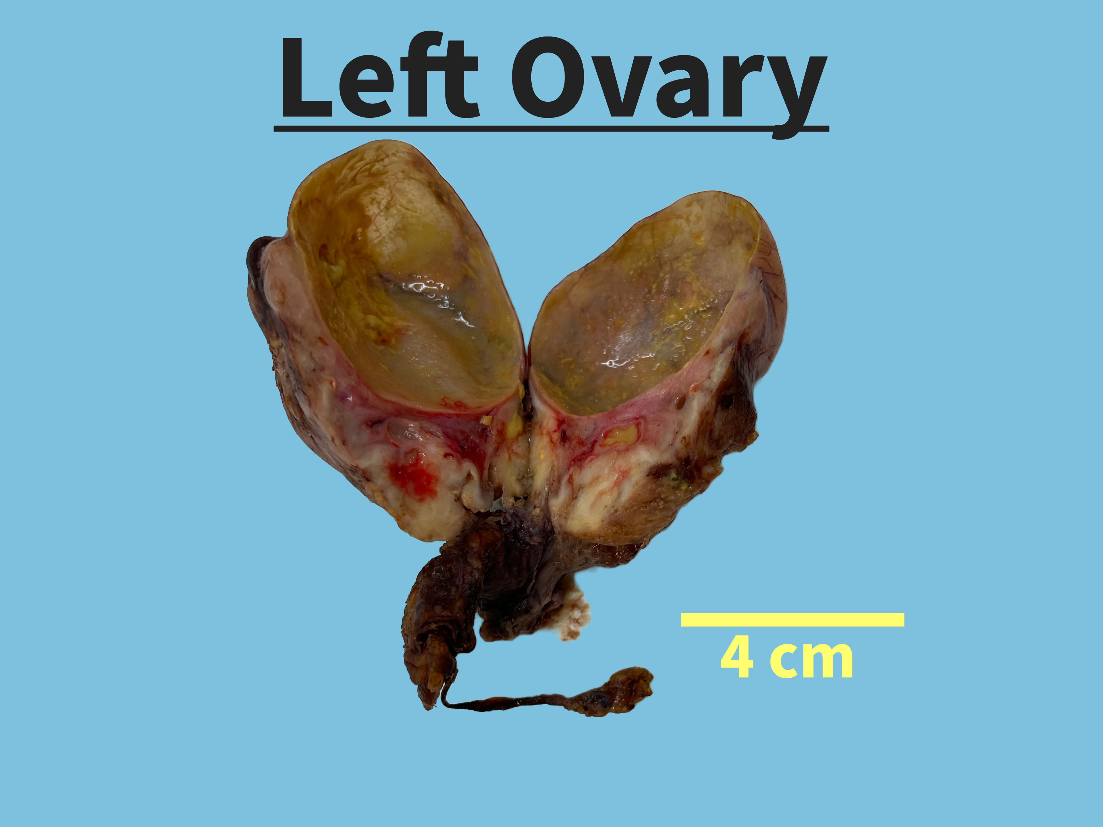
The left ovary (5.5 x 5.0 x 3.9 cm) has a tan-white nodular external surface. Sectioning of the left ovary reveals a softened solid-cystic mass occupying the entirety of the cut surface. The mass is comprised of yellow-white nodular areas (40%) and multiloculated cyst (60%) ranging from 0.2 cm to 5.0 cm in greatest dimension with yellow fluid and cyst wall thickness measuring up to 0.1 cm. No excrescences are identified. No ovarian parenchyma is identified.
The left fimbriated fallopian tube (3.5 cm in length, diameter ranging from 0.4 to 1.1 cm) has purple-tan serosa remarkable for multiple paratubal cysts ranging from 0.2 to 0.3 cm in greatest dimension with cloudy contents. Sectioning reveals a lumen ranging from pinpoint to stellate.
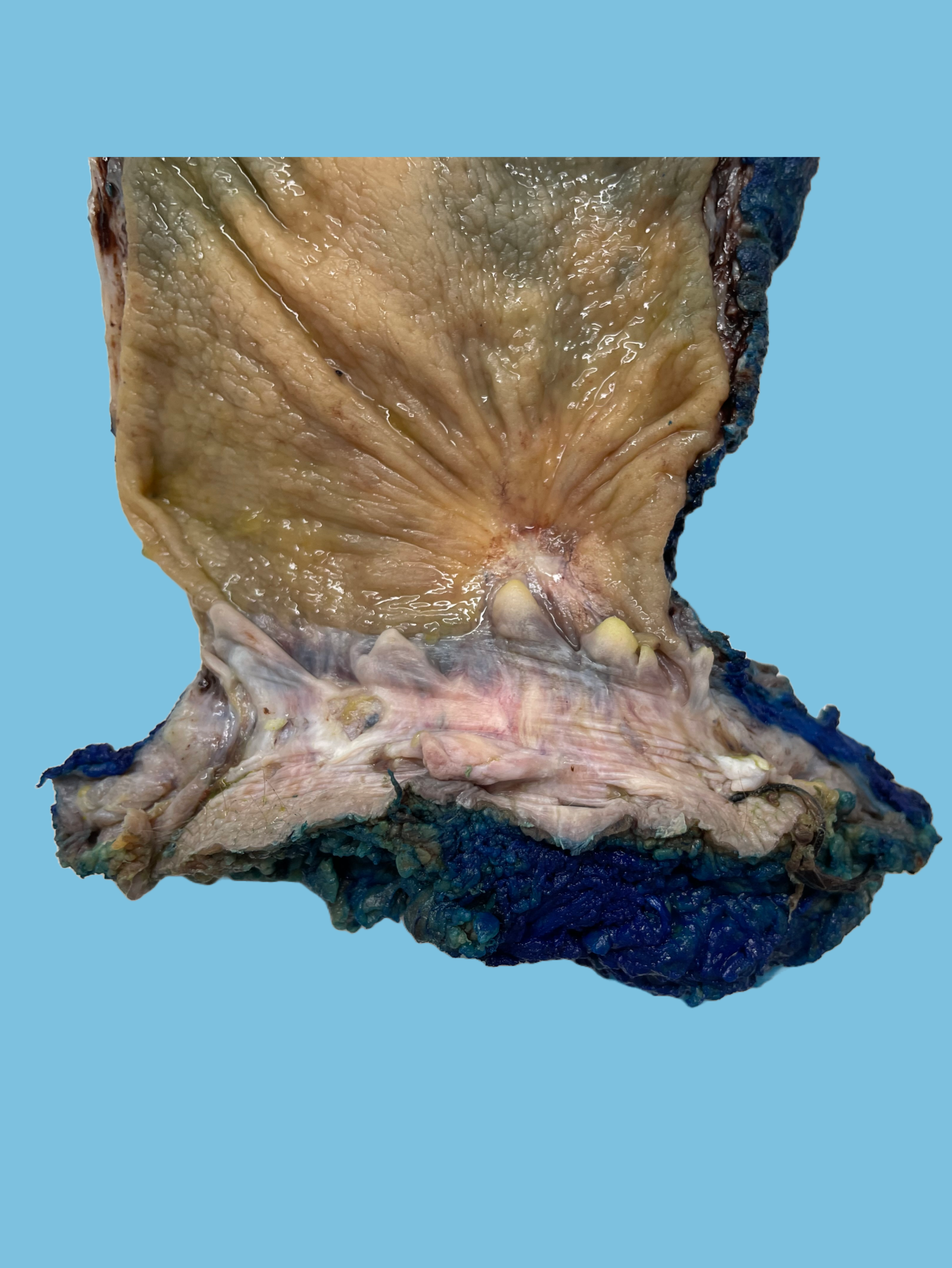
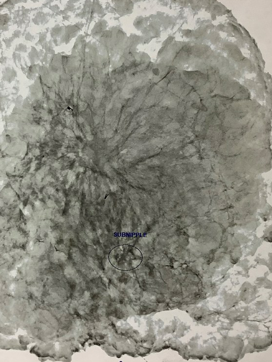
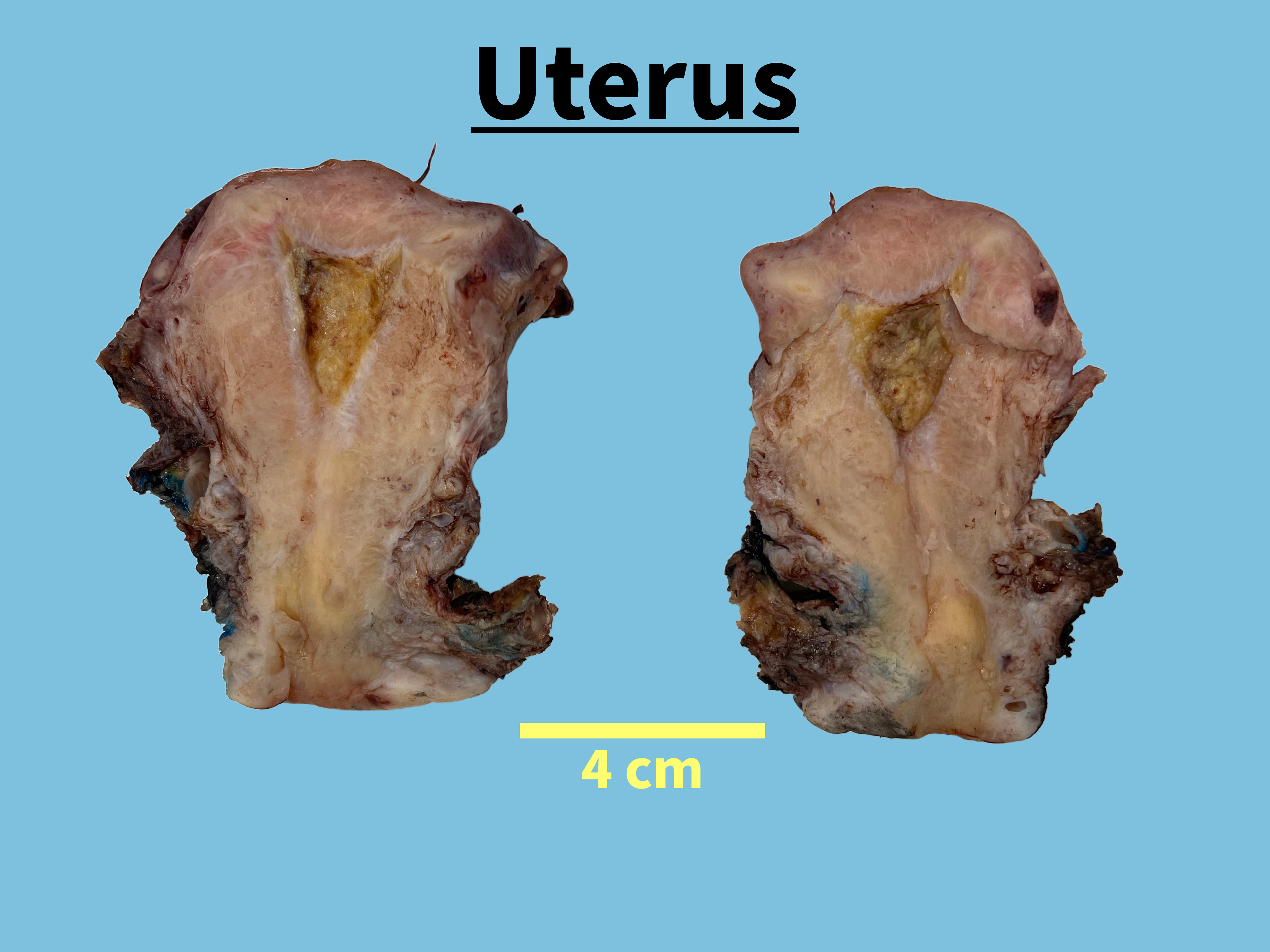
The uterus (8.5 cm from fundus to cervix, 5.5 cm from cornu to cornu, 4.3 cm from posterior to anterior) has a tan-pink serosa remarkable for multiple tan-white subserosal nodules ranging from 0.5 to 2.1 cm in greatest dimension. The cervix (3.8 x 3.7 cm) has tan-white ectocervical mucosa with areas of petechiae, and an ovoid cervical os (1.2 cm). Attached to the right cervix is tan-pink paracervical soft tissue measuring 3.0 x 2.5 x 1.5 cm. A scant amount of pale-pink parametrium is present. The specimen is inked as follows: anterior lower uterine segment–blue, posterior lower uterine segment– black. The uterus is bivalved to reveal an endocervical canal measuring 2.1 cm in length, with a diameter ranging from 0.1 to 0.9 cm. The endometrial cavity (1.9 cm from cornu to cornu, 2.6 cm from fundus to lower uterine segment) is pale yellow, diffusely cauterized and is remarkable for friable contents occupying the entire cavity. Normal endometrium is not identified. There is a fibrous stenosis of the lower uterine segment (0.1 cm). The myometrium is tan-pink, and a shows multiple intramural nodules and subserosal nodules ranging from 0.2 to 2.1 cm with white whorled bulging cut surfaces. The largest subserosal nodule is remarkable for an area of calcification (40%). No additional masses, lesions or abnormalities are identified.
Multiple gross photographs are taken. Representative sections are submitted as follows:
D1: Left Parametrium margin, enface (posterior inked black)
D2-D3: Left parametrial soft tissue with potential mass, en face
D4: Right parametrial margin, en face (posterior inked black)
D5: Posterior right paracervical margin, en face
D6: Anterior right paracevical margin, en face
D7-D8: Lower uterine segment to cervix (12:00)
D9-D10: Lower uterine segment to cervix (6:00)
D11: Anterior lower uterine segment with fibrous stenosis, full-thickness
D12: Posterior uterine body, full-thickness with friable contents and nodule
D13: Nodules, serosa with potential mass
D14: Left fallopian tube with potential mass, fimbriae (bisected)
D15-D19: Left Ovary
D15-D16: Mass, solid areas
D17-D19: Mass, solid to cystic areas
D20: Right fallopian tube, fimbriae (bisected)
D21: Right broad ligament with potential mass
D22-D28: Right ovary
D22-D23: Mass extension through capsule
D24-D25: Papillary excrescences
D26: Cystic areas
D27: Nodular solid areas to cystic areas
D28: Nodular solid areas