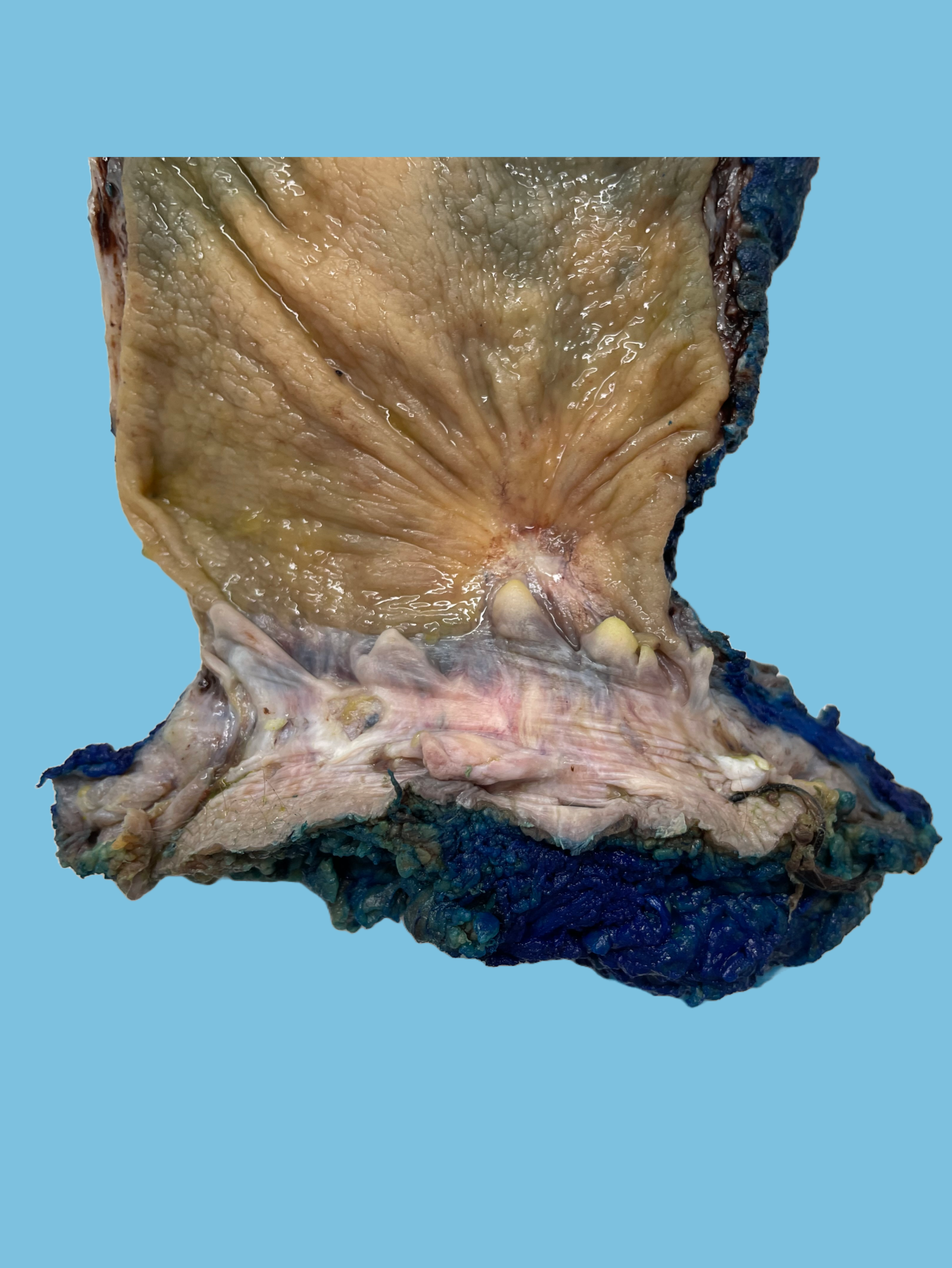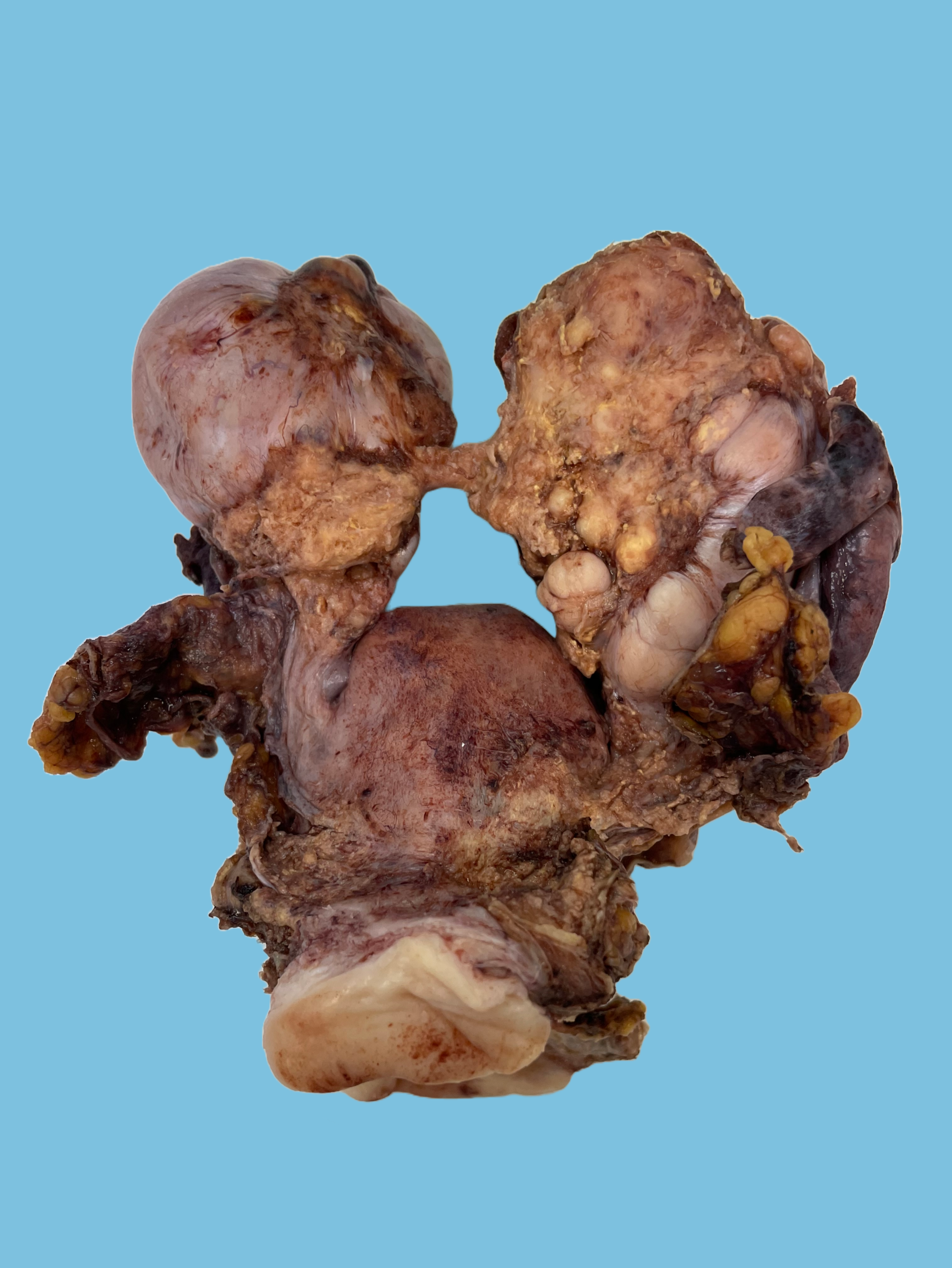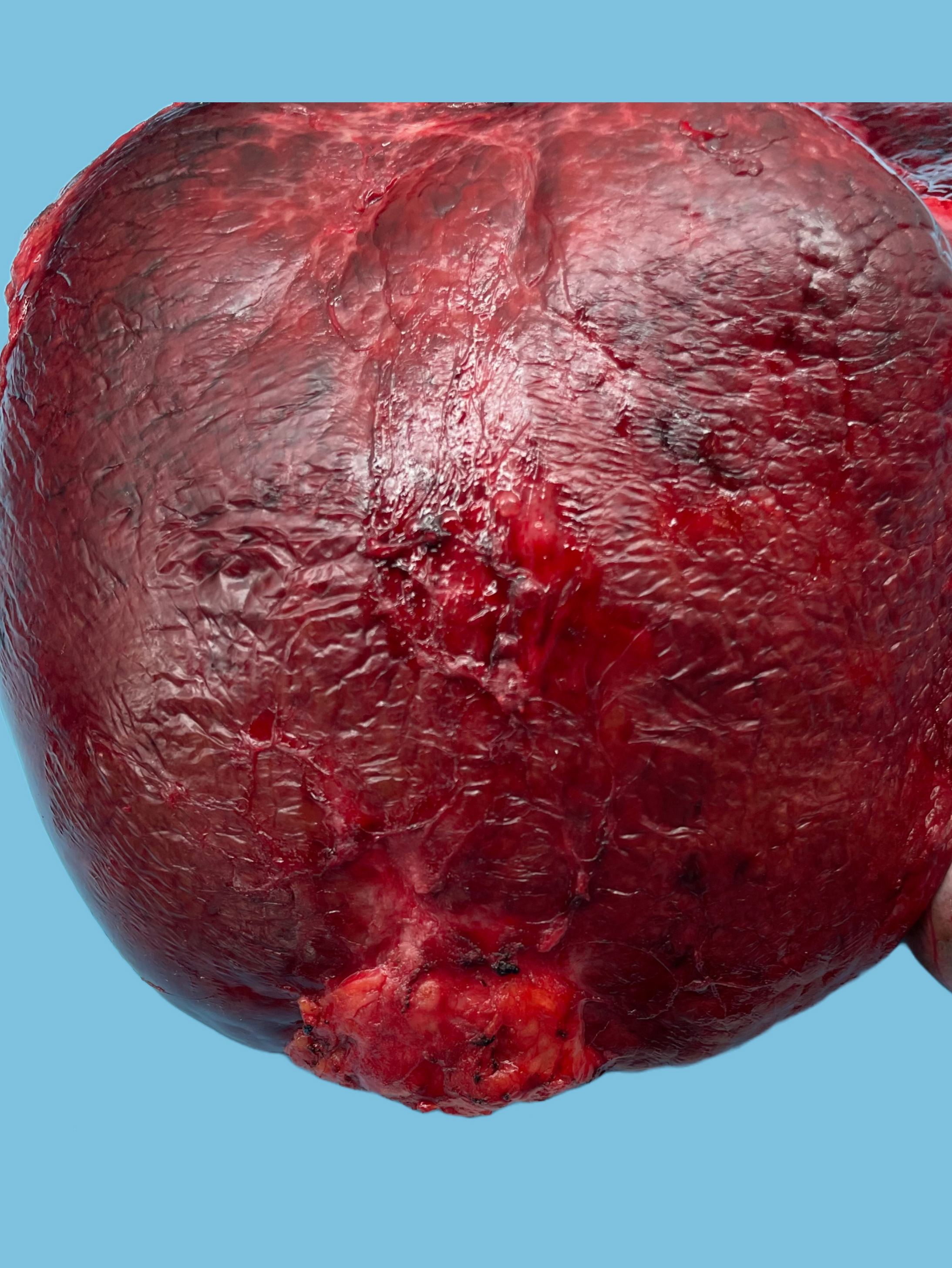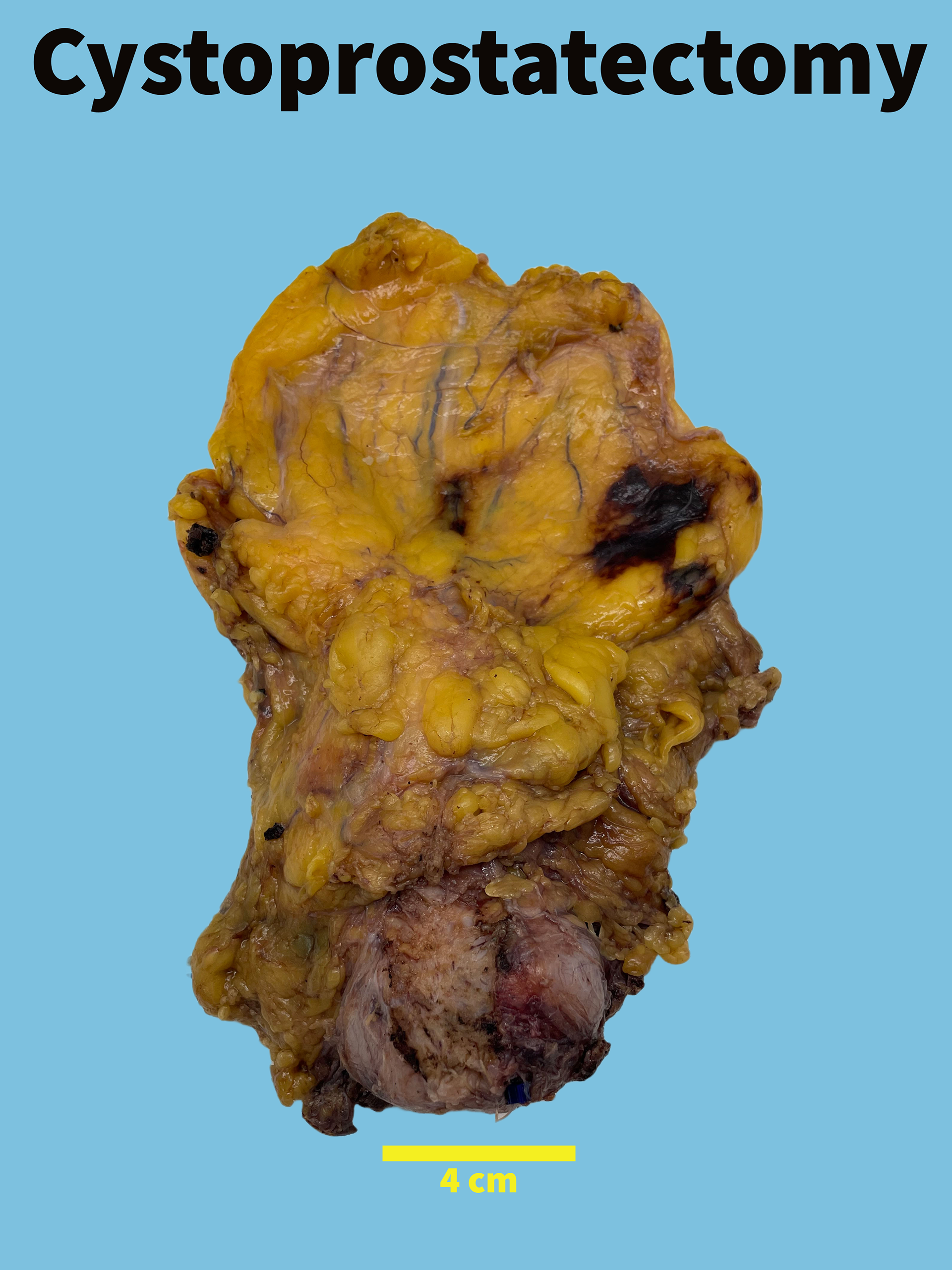
Received fresh for routine processing and subsequently placed in formalin by the Surgical Pathology Department, labeled with the patient's ID and labeled on the container " bladder, prostate, and seminal vesicles" is an intact cystoprostatectomy consisting of bladder (7.0 x 6.0 x 4.2 cm), attached right ureter (1.0 cm in length, 0.5 cm in diameter), left ureter (1.2 cm in length, 0.6 cm in diameter), prostate (4.2 cm from apex to base, 5.2 cm from left to right, and 3.0 cm from posterior to anterior), right vas deferens (8.0 cm in length, 0.5 cm in diameter), left vas deferens 7.0 cm in length, 0.4 cm in diameter), possible right seminal vesicle (1.5 x 0.6 x 0.3 cm), left seminal vesicle (2.8 x 1.0 x 0.7 cm). The bladder serosal surface is tan-yellow, smooth and unremarkable. The perivesical soft tissue margin is roughened without disruptions. The specimen is inked as follows: Right bladder margin and right anterior prostate–green, left bladder margin and left anterior prostate–orange, right posterior soft tissue margin and right posterior prostate– black, left posterior soft tissue margin and left posterior prostate-blue.
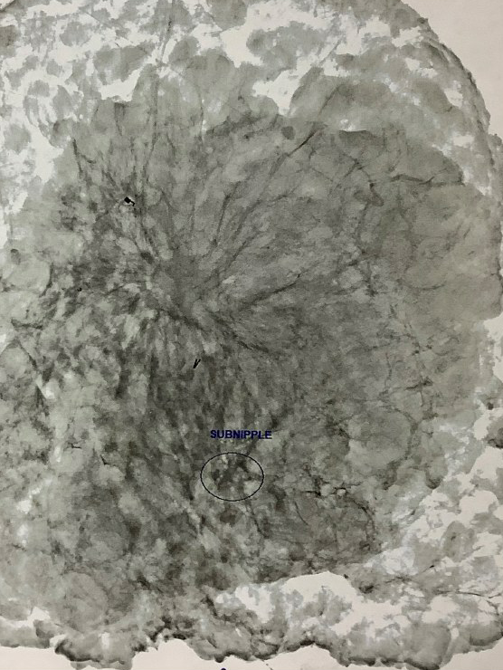
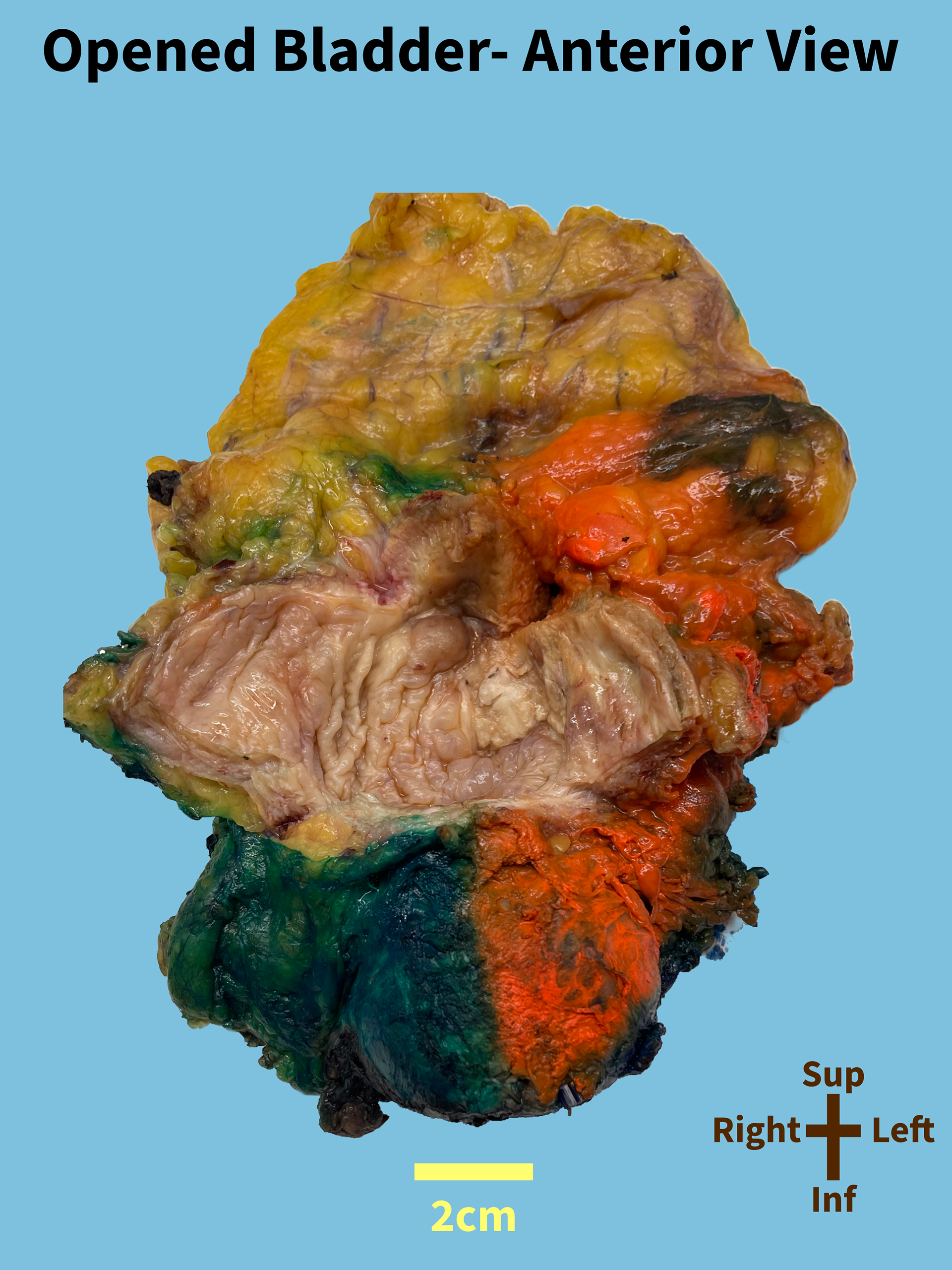
The bladder is opened to reveal a 3.2 x 3.0 cm tan-white grainy fibrous area occupying a portion of the posterior wall, left lateral wall, dome, and anterior wall. Sectioning reveals the fibrous area confined to the mucosa. The fibrous area is 1.0 cm from the serosa, 1.2 cm from the left ureteric orifice 2.1 cm from the right ureteric orifice, 3.2 cm to the bladder neck, 3.6 cm to the prostate, 4.2 cm to the left ureter margin, 5.0 cm right ureter margin, and greater than 6.0 cm to the urethral margin and vasa deferentia margins.
Additionally identified are focal edematous areas on the posterior wall, trigone area, right lateral wall. The remaining mucosa is tan-white with a normal folding pattern, and a transmural thickness of 0.6 cm. The ureters are opened to reveal pale-tan mucosa. No masses or lesions are identified.
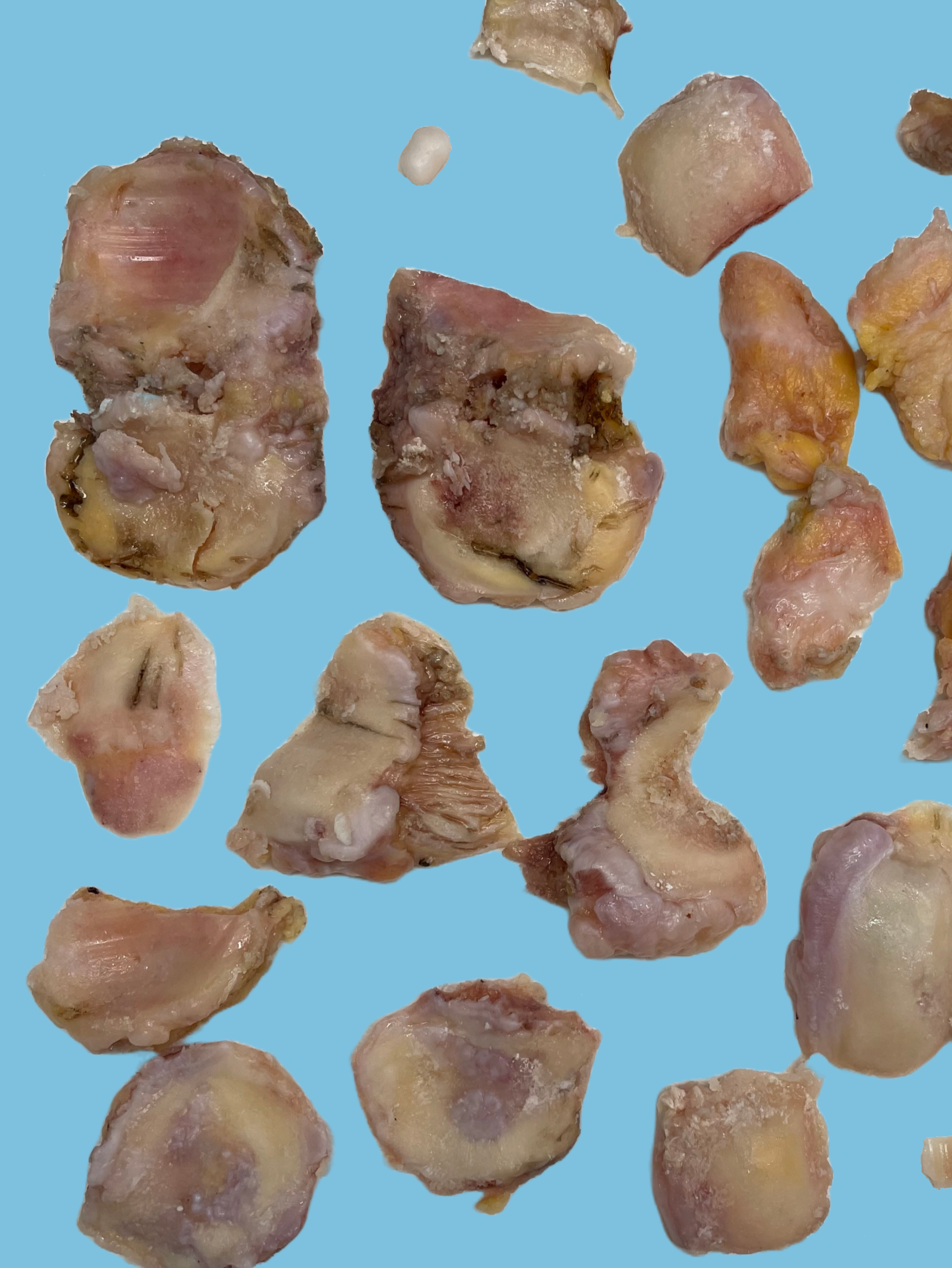
The prostate has a tan-pink smooth posterior surface, and a tan-pink roughened anterior surface with a 2.5 x 2.0 cm area of cautery. The prostate is serially sectioned from base to apex to base into 9 slices to reveal tan-pink to tan-gray periurethral nodularity. Additionally identified are 3 yellow softened areas (ranging from 0.3 to 0.5 cm) in the left posterior aspect of slice 5, and the left anterior aspect of slice 6 and 7. Sectioning of the vasa deferentia and seminal vesicle reveal unremarkable cut surfaces. No masses or lesions are identified.
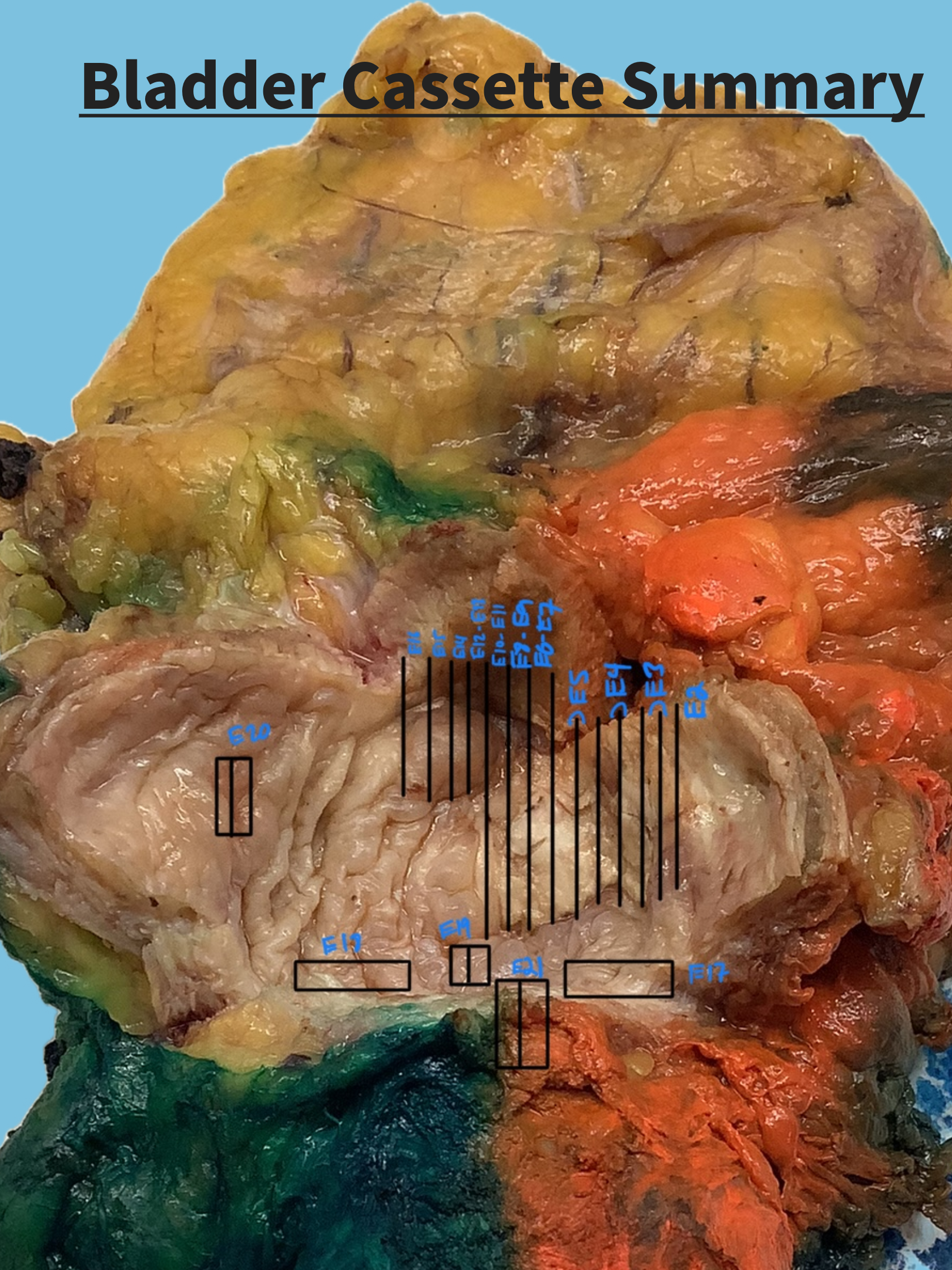
Multiple gross photographs, including an annotated gross photograph are taken. Representative sections to include the entire fibrous area are submitted as follows:
Multiple gross photographs, including an annotated gross photograph are taken. Representative sections to include the entire fibrous area are submitted as follows:
E1: Bilateral ureteral margins (right-inked black, left-inked blue), shave
E2-E16: Fibrous area blocked out
E2-E5: Left lateral wall,
E2: tissue lateral to fibrous area, full thickness
E6–: Posterior wall to dome to anterior
E6-E7: Posterior to anterior, bisected
E8-E9: Posterior to anterior full thickness, fibrous area to serosa and anterior margin, bisected
E10–E11: Posterior to anterior, trisected
E12–E13: Posterior to anterior, bisected
E12: Edematous posterior wall
E14-E16: Posterior to anterior
E17: Left ureter orifice
E18: Right ureter orifice
E19: Edematous trigone area
E20: Right lateral wall
E21: Bladder neck
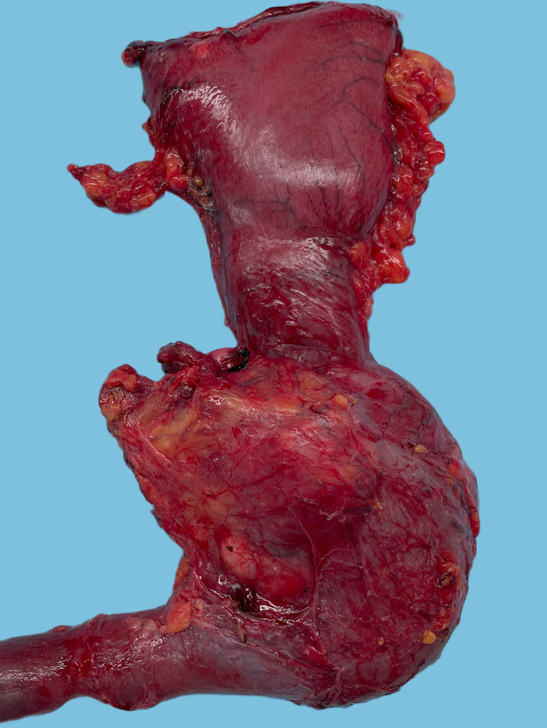
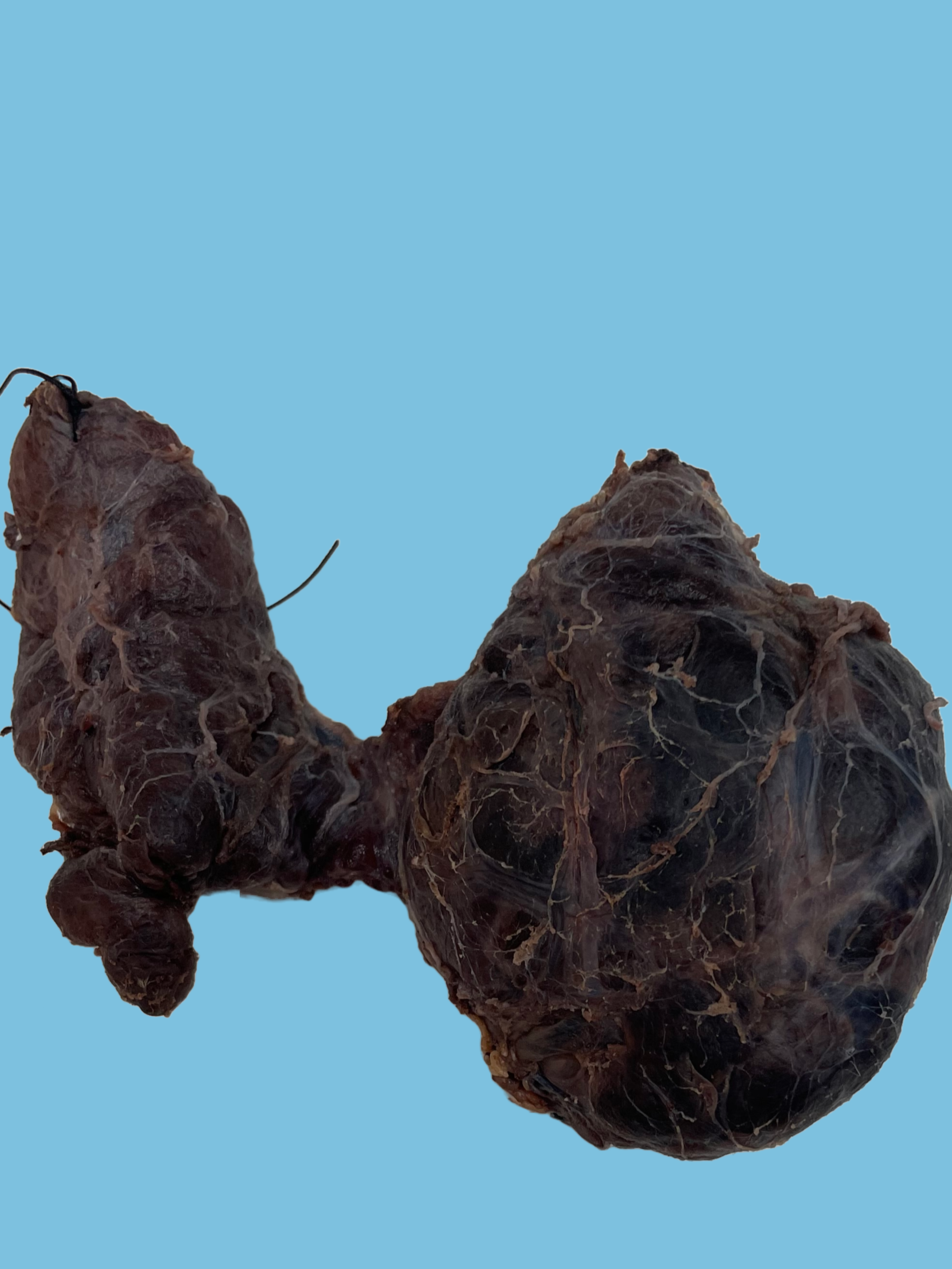
E22: Right seminal vesicle, right vas deferens, right vas deferens margin (shave)
E23: Left seminal vesicle, left vas deferens, right and left vas deferens margin (shave)
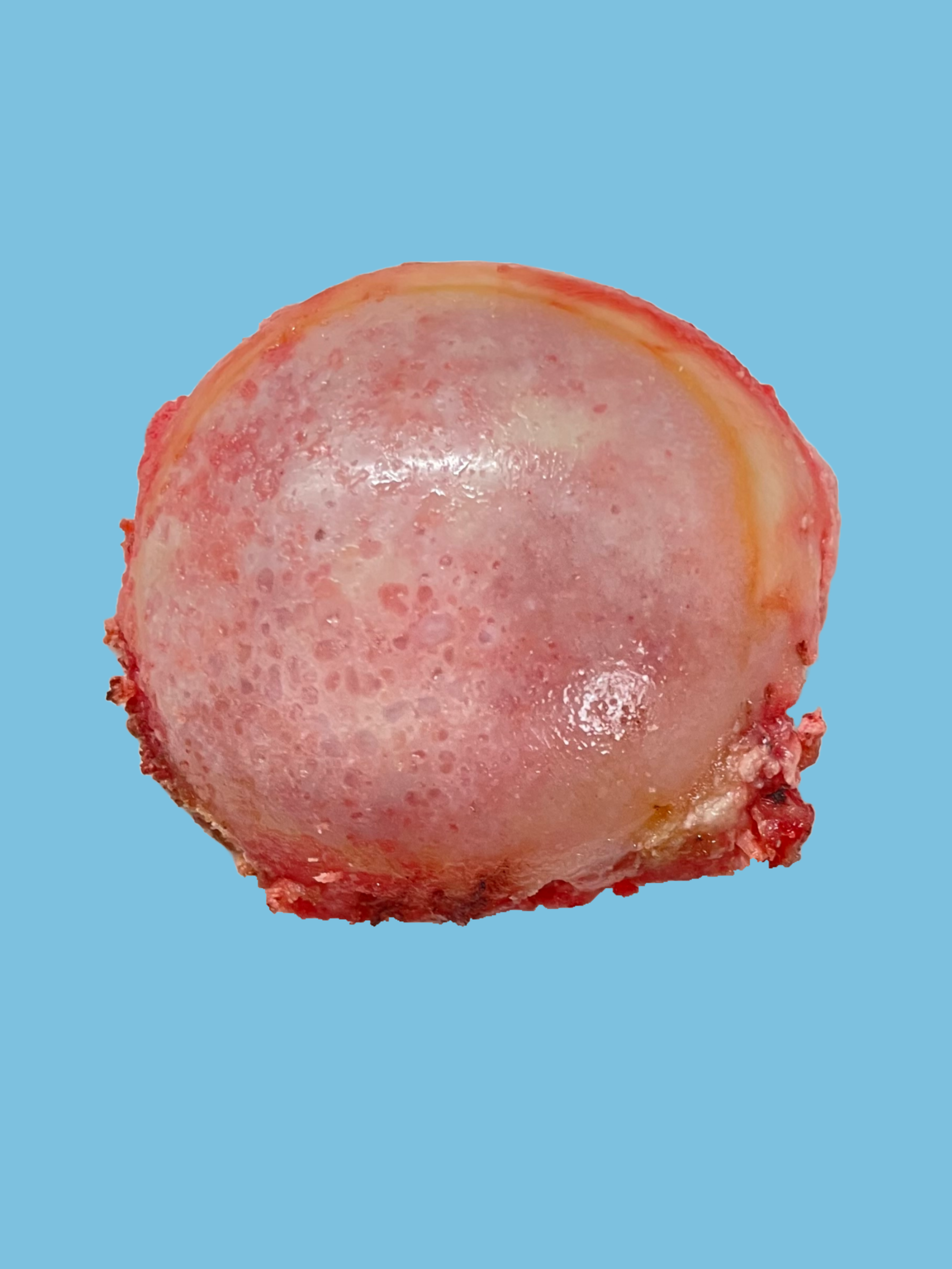
E24: Slice 9, cystic area
E25-E26: Slice 8, lateral aspects
E27: Slice 7, yellow softened area in left anterior aspect
E28–E30: Slice 6
E28: Yellow softened area in left anterior aspect
E29-E30: Lateral aspects
E31–E33: Slice 5, posterior half, trisected
E31: Yellow softened area in left posterior aspect
E34–E35: Slice 3, entirely, bisected
E36–E37: Slice 2, entirely, bisected
E38-E39: Slice 1 entirely, apical margin, perpendicular
E40–E49: Perivesicular adipose tissue